Lighthouse Health Education Center
Click on a blue query hyperlink below or enter a keyword to see research and resources about that keyword.

Near-infrared light increases ATP, extends lifespan and improves mobility in aged Drosophila melanogaster

Rana Begum 1 , Karin Calaza 2 , Jaimie Hoh Kam 1 , Thomas E. Salt 1 , Chris Hogg 3 and Glen Jeffery - Royal Society Publishing (Publication)
PBM increased the average lifespan and mobility of fruit flies. Although they all died at 12 weeks, treating the flies with PBM significantly increase the average healthspan.
View Resource

Near-infrared light increases ATP, extends lifespan and improves mobility in aged Drosophila melanogaster Rana Begum 1 , Karin Calaza 2 , Jaimie Hoh Kam 1 , Thomas E. Salt 1 , Chris Hogg 3 and Glen Jeffery Institute of Ophthalmology, University College London, London EC1V 9EL, UK 2 Program of Neuroscience, Institute de Biologia, Universidade Federal Fluminense, Rio de Janeiro 24210130, Brazil 3 Moorfields Eye Hospital, London EC1V 2PD, UK Ageing is an irreversible cellular decline partly driven by failing mitochondrial integrity. Mitochondria accumulate DNA mutations and reduce ATP production necessary for cellular metabolism. This is associated with inflammation. Near-infrared exposure increases retinal ATP in old mice via cytochrome c oxidase absorption and reduces inflammation. Here, we expose fruitflies daily to 670 nm radiation, revealing elevated ATP and reduced inflam- mation with age. Critically, there was a significant increase in average lifespan: 100–175% more flies survived into old age following 670 nm exposure and these had significantly improved mobility. This may be a simple route to extending lifespan and improving function in old age. 1. Introduction Mitochondria provide cellular energy via adenosine triphosphate (ATP). But, their DNA (mtDNA) suffers from progressive mutations resulting in reduced ATP production, which is thought to run concomitantly with an increase in pro-inflammatory reactive oxygen species (ROS) [1,2]. Hence, hallmarks of ageing are reduced cellular energy and progressive systemic inflammation. Meta- bolic demand also plays a role as tissues and organisms with high metabolic rates generally suffer from rapid ageing [3,4]. The retina has the greatest metabolic demand in the body [5], but ATP decline in the central nervous system can be significantly improved by near-infrared/infrared light (NIR/IR, [6]). Specific wavelengths in this range are absorbed by cytochrome c oxidase in mitochondrial respiration, improving its efficiency [7–10]. These wavelengths improve mito- chondrial membrane potentials, significantly reduce inflammation and reduce macrophage numbers with brief exposures of around 60–90 s repeated over approximately a week [11,12]. NIR/IR also reduces experimental pathology when insult impacts on mitochondrial function, as in experimental Parkinson’s disease, where NIR significantly reduces cell death in the substantia nigra [13]. However, NIR/IR studies have largely used light for short periods and their impact on lifespan has not been assessed [7,11,12]. If NIR improves mitochondrial function we predict it may extend life. The fly has been used here because of its relatively short life [14]. Hence, we ask if long-term exposure to 670 nm in Drosophila melanogaster can increase lifespan and improve function in old age. 2. Material and methods Drosophila melanogaster were used. Hatched male flies were housed on 12/12 light cycle at 258C within a season. Half were exposed to 670 nm for 20 min per day at & 2015 The Authors. Published by the Royal Society under the terms of the Creative Commons Attribution License http://creativecommons.org/licenses/by/4.0/, which permits unrestricted use, provided the original author and source are credited. on March 18, 2015http://rsbl.royalsocietypublishing.org/Downloaded from 40 mW cm 22 in clear plastic 50 cm 3 (28 mm wide) containers, illuminating flies from either side, which were counted weekly. Room illumination was 2 mW cm 22 . 670 nm energies were approximately 100 times lower than indirect sunlight, consistent with earlier studies [7]. Light devices were built by C. H. Elec- tronics UK and contained 50 670 nm LEDS over 20 cm 2 . Six independent replicates were used in lifespan experiments (n ¼ 620 flies). ATP, inflammation and mobility were assessed at seven weeks, when ATP and mobility are known to decline [15]. ATP was measured by luciferin–luciferase assay (Enliten w ATP Assay System, Promega). Flies were killed with liquid nitrogen, transferred to 2.5% trichoroacetic acid (TCA), then homogenized at 48C. Supernatant was collected and the TCA was neutralized with 1 M Tris–acetate buffer (pH 7.75, final TCA concentration 0.0625%); 10 ml of neutralized solution was added to 100 ml of luci- ferin–luciferase in fresh buffer. ATP was measured using an Orion microplate luminometer (Berthold Detection Systems GmbH) and data normalized to fly numbers. Tissues were homogenized in 2% sodium dodecyl sulfate (SDS) with protease inhibitor cocktail for Western blot (Roche Diagnos- tics), and centrifuged; the supernatant was pipetted out, separated with 10% SDS–PAGE and electrophoretically transferred onto nylon membranes. Immunoblotting was undertaken for complement component C3 (Cappel, MP Biomedicals), which is highly conserved [16]. Protein was quantified by densitometric X-ray scanning and values were normalized to a-tubulin. Fly mobility assessment was as Bjedov et al. [14]. Flies were placed in 100 ml clear cylinders (seven flies per trial), tapped to the bottom and then videoed, the last two steps repeated three times. Using the videos, the number of flies above the 50 ml mark (9 cm from the bottom) was counted after 1 min. Individual flies were traced, with absolute distance travelled measured. Data w er e analysed with GraphPad P RISM v. 5 and sta t is ti cal analysis was undertaken using Mann –Whitne y U non-parametric and log-rank tests. 3. Results (a) ATP levels are elevated and systemic inflammation reduced Whole body ATP declines with age only after appr o xima tely seven weeks [14], when ATP was measured here. A TP concen- trations wer e significantly grea t er, by approxima tely 80%, in 670 nm exposed animals compared with unexposed (figure 1a, Mann–Whitney test p ¼ 0.028). At seven weeks, W estern blots wer e undertaken for inflammatory marker complement com- ponent C3. This was reduced in 670 nm exposed flies compared with controls (figure 1b). Hence, 670 nm radiation elevates ATP and reduces inflammation. (b) Lifespan increases Fly numbers in experimental and control groups were similar in the two weeks post-hatching. From week 3, fly deaths were greater in controls than 670 nm exposed flies and they remained so at each time point until week 11–12, when all flies were dead in both groups. This difference was significant (figure 2, log-rank test p ¼ 0.008). The progressive mean percentage increase in 670 nm flies alive over controls is given in figure 2b. Group differences accelerated from week 4, when 10% extra 670 nm treated flies were alive compared with controls, to approximately 50% extra when the control population had halved. By the time the control population was reduced by 80%, at week 8, more than 100% extra 670 nm treated flies remained alive. Subsequently, group differences reached almost 180% before declining to zero in both groups at week 11–12. Hence, 670 nm did not extend absolute lifespan. (c) Aged mobility increases Mobility of 670 nm treated and control flies was measured at seven weeks. Significantly more 670 nm treated flies climbed above the 50 ml level (9 cm) and significantly more travelled a greater distance than controls (Mann–Whitney test p ¼ 0.028, p ¼ 0.014, respectively). Twice as many 670 nm flies climbed above 50 ml (9 cm) compared with controls and these travelled twice the distance in 1 min compared with controls (figure 2c,d). Hence, 670 nm exposure significantly improves both lifespan and mobility. 4. Discussion Drosophila melanogaster has been widely used in lifespan studies as they are short lived and their genomic sequence is relatively well understood [14,17], hence their adoption experimentally here to extend lifespan. Our results reveal [ATP] (nM)/fly ATP level(a) C3 expression (Western blot)(b) control absolute intensity 670 control 670 control C3 ~ 110 kDa a-tubulin 55 kDa 670 * 0 0 500 1000 200 400 600 Figure 1. Exposur e to 670 nm radiation increases ATP in aged flies and reduces inflammati on. (a) Sev en week old flies exposed to 670 nm had a significant incr ease in whole body ATP compared with controls, p ¼ 0.028. n ¼ 25 flies per group. (b) Whole body inflammation (C3) was measured in seven week flies using W estern blot. This was reduced in 670 nm exposed flies by approxima tely 15%. Here, flies were pooled within groups as C3 protein levels were lo w in individuals. Hence there are no error bars. n ¼ 15 flies per group. (Online version in colour.) rsbl.royalsocietypublishing.org Biol. Lett. 11: 20150073 2 on March 18, 2015http://rsbl.royalsocietypublishing.org/Downloaded from that when flies are exposed to 670 nm radiation they have reduced inflammation, improved ATP, improved mobility and extended average lifespans. These data are consistent with the majority of studies undertaken using 670 nm on mammals, showing reduced inflammation in experimental models and in ageing, and improved ATP levels [6,7]. How- ever, it would be difficult to undertake lifespan experiments in mice as the light would not penetrate the entire body as it does in flies and hence its influence would not be systemic. There are many factors and pathways in ageing, and nine candidate hallmarks have been suggested, which may be separate, but also are likely to have interactions [1]. Mito- chondrial function is one. Previously, mitochondrial function and ageing were viewed within a framework of pro- gressive mtDNA mutations/deletions resulting in reduced ATP and increased ROS. The balance of these factors was seen as a driver in the mitochondrial theory of ageing [18]. However, evidence has undermined the role of ROS in ageing [19,20]. Hence, some mutant mice have reduced life- span as a result of mtDNA mutations/deletions not associated with increased ROS [21,22]. Further, increased ROS can prolong lifespan in yeast and Caenorhabditis elegans [22,23], and in mammals it does not accelerate ageing [20]. These data are reviewed by Lopez-Otin et al. [1], who argue that low ROS may activate compensatory mechanisms and not directly contribute to ageing. Such data may undermine the ROS element in Harman’s mitochondrial theory [18]. If correct, it places greater potential emphasis on ATP in ageing. NIR has been successful in treating induced pathology [7] and ageing, particularly in the retina, where progressive age- related inflammation is marked owing to high metabolic rate [11,12]. These wavelengths penetrate deeply and 670 nm trans-illuminated our flies at 40 mW cm 22 . In relation to this, it may be significant that, while old domestic incandescent light- ing contained significant NIR elements, none is present in modern strip lighting or energy-saving domestic lighting [12]. The absence of these wavelengths from artificial lighting may have long-term consequences. As longer wavelengths penetrate deeply, this may be of significance not only for the ageing eye, but also potentially for other tissues. Ethics statement. Fly research is free of legal ethical constraint. Data accessibility. All data are presented in the manuscript. Acknowledgement. We thank Iris Salecker, Giovanna Vinti and Tobi Weinrrich for technical assistance. Author contributions. G.J. designed experiments and wrote the manu- script. All authors undertook the experiments and approved the final version of the manuscript. R.B. analysed the data. Funding statement. Supported by the Rosetrees Trust UK. K.C. was a research fellow from CAPES Brazil (proc. 18134/12-2). Competing interests. We have no competing interests. References 1. Lopez-Otin C, Blasco MA, Partridge L, Serrano M, Kroemer G. 2013 The hallmarks of aging. Cell 153, 1194–1217. (doi:10.1016/ j.cell.2013.05.039) 2. Balaban RS, Nemoto S, Finkel T. 2005 Mitochondrial, oxidants and aging. Cell 120, 483–495. (doi:10.1016/j.cell.2005.02.001) 3. Speakman JR. 2005 Body size, energy, metabolism and lifespan. J. Exp. Biol. 208, 1717–1730. (doi:10. 1242/jeb.01556) 4. Wang Z, Ying Z, Bosy-Westphal A, Zhang J, Schautz B, Later W, Heymsfield SB, Mu¨ller MJ. 2010 Specific metabolic rates of major organs and tissues across adulthood: evolution by mechanistic model of resting expenditure. Am. J. Clin. Nutr. 92, 1369– 1377. (doi:10.3945/ajcn.2010.29885) 5. Yu DY, Cringle SJ. 2001 Oxygen distribution and consumption within the retina in vascularized and avascular retinas and in animal models of disease. Prog. Retin. Eye Res. 20, 175–208. (doi:10.1016/ S1350-9462(00)00027-6) 0 0 10 20 30 40 50 60 70 80 100 (a) (c) (d ) (b) % survival over controls n = 620 flies fly survival curves for 670 nm (–) and control (–) 90 123456 time (weeks) % survival 78910 0 0 100 200 300 123456 no. weeks % increase over control control distance travelled in 1 min 670 control 670 * 0 20 60 40 80 100 distance (mm) fly climbing (>90 mm) * 0 20 60 40 80 % >90 mm in 1 min 78910 11 12 Figure 2. Lifespan and mobility. (a) Fly numbers at progressive weeks in groups exposed to 670 nm supplemented light each day (red line) and controls (black line). Curves are averages for six independent experiments with a minimum of 40 flies per group in each experiment. Fly death rates separated between three and six weeks with fewer flies dying in 670 nm exposed animals. Reduction in the two population followed similar patterns from six weeks but with the 670 nm exposed group having greater numbers at any point until week 12. In all replicates, there was no indication that 670 nm increased absolute lifespan beyond weeks 11 – 12. Differences between the two groups were statistically significant ( p ¼ 0.008). (b) Inset: percentage increase of 670 nm exposed flies alive at pro- gressive weeks. (c) Seven week old 670 nm exposed flies were more active than controls. (d) Mobility measures the percentage of flies that climbed above 90 mm in a clear 100 ml cylinder. (d) This was filmed and then the distance travelled by each fly was measured in each group. In both cases, the 670 nm exposed flies where significantly more mobile. There were 21 flies in each group in each condition. (Online verion in colour.) rsbl.royalsocietypublishing.org Biol. Lett. 11: 20150073 3 on March 18, 2015http://rsbl.royalsocietypublishing.org/Downloaded from 6. Gkotsi D, Begum R, Salt T, Lascaratos G, Hogg C, Chau KY, Schapira AH, Jeffery G. 2014 Recharging mitochondrial batteries in old eyes. Near infra-red increases ATP. Exp. Eye Res. 122, 50 –53. (doi:10. 1016/j.exer.2014.02.023) 7. Fitzgerald M et al . 2013 Red/near-infrared irradiation therapy for treatment of central nervous system injuries and disorders. Rev. Neurosci. 24, 205–226. (doi:10.1515/revneuro-2012-0086) 8. Wilson M, Greenwood C. 1970 The long-wavelength absortion band of cytochrome c oxidase. Biochem. J. 116, 17 –18. 9. Karu TI, Pyatibrat LV, Kolyakov SF, Afanasyeva NI. 2005 Absorption measurements of cell monolayer relevant to phototherapy: reduction of cytochrome c oxidase under near IR radiation. J. Photochem. Photobiol. 81, 98– 106. (doi:10.1016/j.jphotobiol.2005.07.002) 10. Cooper CE, Springett R. 1997 Measurement of cytochrome oxidase and mitochondrial energetics by near-infrared spectroscopy. Phil. Trans. R. Soc. Lond. B 352, 669– 676. (doi:10.1098/rstb.1997.0048) 11. Kokkinopoulos I, Colman A, Hogg C, Heckenlively J, Jeffery G. 2013 Age-related inflammation is reduced by 670 nm light via increased mitochondrial membrane potential. Neurobiol. Aging 34, 602– 609. (doi:10.1016/j.neurobiolaging.2012.04.014) 12. Begum R, Powner MB, Hudson N, Hogg C, Jeffery G. 2013 Treatment with 670 nm up regulates cytochrome C oxidase expression and reduces inflammation in an age-related macular degeneration model. PLoS ONE 8, e57828. (doi:10. 1371/journal.pone.0057828) 13. Purushothuman S, Nandasena C, Johnstone DM, Stone J, Mitrofanis J. 2013 The impact of near- infrared light on dopaminergic cell survival in a transgenic mouse model of parkinsonism. Brain Res. 1535, 61 –70. (doi:10.1016/j.brainres.2013.08.047) 14. Bjedov I, Toivonen JM, Kerr F, Slack C, Foley A, Partridge L. 2010 Mechanisms of life span extension by rampamycin in the fruit fly Drosophila melanogaster. Cell Metab. 11, 35–46. (doi:10.1016/ j.cmet.2009.11.010) 15. Vernace VA, Arnaud L, Schmidt-Glenewinkel T, Figueiredo-Pereira ME. 2007 Aging perturbs 26S proteasome assembly in Drosophila melanogaster . FASEB J. 21, 2672–2682. (doi:10.1096/fj.06- 6751com) 16. Nonaka M, Kimura A. 2006 Genomic view of the evolution of the complement system. Immunogenetics 58, 701– 713. (doi:10.1007/ s00251-006-0142-1) 17. Celniker SE, Rubin GM. 2003 The Drosophila melanogaster genome. Annu. Rev. Genomics Hum. Genet. 4, 89–117. (doi:10.1146/annurev.genom.4. 070802.110323) 18. Harman D. 1981 The ageing process. Proc. Natl Acad. Sci. USA 78, 7124 –7128. (doi:10.1073/pnas. 78.11.7124) 19. Edgar D et al. 2009 Random point mutations with major effects on protein coding genes are the driving force behind premature aging in mtDNA mutator mice. Cell Metab. 10, 131–138. (doi:10. 1016/j.cmet.2009.06.010) 20. Hiona A et al. 2010 Mitcohondrial DNA mutations induce mitochondrial dysfunction, apoptosis and sarcopenia in skeletal muscle of mitochondrial mutator mice. PLoS ONE 5, e11468. (doi:10.1371/ journal.pone.0011468) 21. Doonan R, McElwee JJ, Matthijssens F, Walker GA, Houthoofd K, Back P, Matscheski A, Vanfleteren JR, Gems D. 2008 Against the oxidative damage theory of aging: superoxide disumatases protect against oxidative stress but have little or no effect on life span in Caenorhabditis elegans. Genes Dev. 22, 3236–3241. (doi:10.1101/gad. 504808) 22. Mesquita A et al. 2010 Caloric restriction or catalase inactivation extends yeast chronological lifespan by inducing H 2 O 2 and superoxide dismutase activity. Proc. Natl Acad. Sci. USA 107, 15 123–15 128. (doi:10.1073/pnas.1004432107) 23. Zhang Y et al. 2009 Mice deficient in both Mn superoxide dismutase and glutathione peroxidase-1 have increased oxidative damage and a greater incidence of pathology but no reduction in longevity. J. Gerontol. A Biol. Sci. Med. Sci. 64, 1212– 1220. (doi:10.1093/gerona/glp132) rsbl.royalsocietypublishing.org Biol. Lett. 11: 20150073 4 on March 18, 2015http://rsbl.royalsocietypublishing.org/Downloaded from


Original Source: https://www.researchgate.net/publication/273781783_Near-infrared_light_increases_ATP_extends_lifespan_and_improves_mobility_in_aged_Drosophila_melanogaster

Effect of NASA light-emitting diode irradiation on wound healing

Whelan HT1, Smits RL Jr, Buchman EV, Whelan NT, Turner SG, Margolis DA, Cevenini V, Stinson H, Ignatius R, Martin T, Cwiklinski J, Philippi AF, Graf WR, Hodgson B, Gould L, Kane M, Chen G, Caviness J. - J Clin Laser Med Surg. 2001 Dec;19(6):305-14. (Publication)
Study showed increases in growth of 155-171% of normal human epithelial cells and an improvment of greater than 40% in musculoskeletal training injuries in Navy SEAL
View Resource

OBJECTIVE:

The purpose of this study was to assess the effects of hyperbaric oxygen (HBO) and near-infrared light therapy on wound healing.

BACKGROUND DATA:

Light-emitting diodes (LED), originally developed for NASA plant growth experiments in space show promise for delivering light deep into tissues of the body to promote wound healing and human tissue growth. In this paper, we review and present our new data of LED treatment on cells grown in culture, on ischemic and diabetic wounds in rat models, and on acute and chronic wounds in humans.

MATERIALS AND METHODS:

In vitro and in vivo (animal and human) studies utilized a variety of LED wavelength, power intensity, and energy density parameters to begin to identify conditions for each biological tissue that are optimal for biostimulation.

RESULTS:

LED produced in vitro increases of cell growth of 140-200% in mouse-derived fibroblasts, rat-derived osteoblasts, and rat-derived skeletal muscle cells, and increases in growth of 155-171% of normal human epithelial cells. Wound size decreased up to 36% in conjunction with HBO in ischemic rat models. LED produced improvement of greater than 40% in musculoskeletal training injuries in Navy SEAL team members, and decreased wound healing time in crew members aboard a U.S. Naval submarine. LED produced a 47% reduction in pain of children suffering from oral mucositis.

CONCLUSION:

We believe that the use of NASA LED for light therapy alone, and in conjunction with hyperbaric oxygen, will greatly enhance the natural wound healing process, and more quickly return the patient to a preinjury/illness level of activity. This work is supported and managed through the NASA Marshall Space Flight Center-SBIR Program.

Read more at: https://pdfs.semanticscholar.org/1f5b/0a4ce02a9c58dfd8531552fd2d2e2f3e701e.pdf

Original Source: https://www.ncbi.nlm.nih.gov/pubmed/11776448

Mechanisms and applications of the anti-inflammatory effects of photobiomodulation

Michael R Hamblin - PMC 2017 Jul 24 (Publication)
Chronic diseases of the modern age involving systemic inflammation such as type II diabetes, obesity, Alzheimer's disease, cardiovascular disease and endothelial dysfunction are again worth investigating in the context of PBM.
View Resource

Abstract

Photobiomodulation (PBM) also known as low-level level laser therapy is the use of red and near-infrared light to stimulate healing, relieve pain, and reduce inflammation. The primary chromophores have been identified as cytochrome c oxidase in mitochondria, and calcium ion channels (possibly mediated by light absorption by opsins). Secondary effects of photon absorption include increases in ATP, a brief burst of reactive oxygen species, an increase in nitric oxide, and modulation of calcium levels. Tertiary effects include activation of a wide range of transcription factors leading to improved cell survival, increased proliferation and migration, and new protein synthesis. There is a pronounced biphasic dose response whereby low levels of light have stimulating effects, while high levels of light have inhibitory effects. It has been found that PBM can produce ROS in normal cells, but when used in oxidatively stressed cells or in animal models of disease, ROS levels are lowered. PBM is able to up-regulate anti-oxidant defenses and reduce oxidative stress. It was shown that PBM can activate NF-kB in normal quiescent cells, however in activated inflammatory cells, inflammatory markers were decreased. One of the most reproducible effects of PBM is an overall reduction in inflammation, which is particularly important for disorders of the joints, traumatic injuries, lung disorders, and in the brain. PBM has been shown to reduce markers of M1 phenotype in activated macrophages. Many reports have shown reductions in reactive nitrogen species and prostaglandins in various animal models. PBM can reduce inflammation in the brain, abdominal fat, wounds, lungs, spinal cord.

2.1. Cytochrome c oxidase in mitochondria

Cytochrome c oxidase (CCO) is unit IV in the mitochondrial electron transport chain. It transfers one electron (from each of four cytochrome c molecules), to a single oxygen molecule, producing two molecules of water. At the same time the four protons required, are translocated across the mitochondrial membrane, producing a proton gradient that the ATP synthase enzyme needs to synthesize ATP. CCO has two heme centers (a and a3) and two copper centers (CuA and CuB). Each of these metal centers can exist in an oxidized or a reduced state, and these have different absorption spectra, meaning CCO can absorb light well into the NIR region (up to 950 nm) [9]. Tiina Karu from Russia was the first to suggest [10,11], that the action spectrum of PBM effects matched the absorption spectrum of CCO, and this observation was confirmed by Wong-Riley et al in Wisconsin [12]. The assumption that CCO is a main target of PBM also explains the wide use of red/NIR wavelengths as these longer wavelengths have much better tissue penetration than say blue or green light which are better absorbed by hemoglobin. The most popular theory to explain exactly why photon absorption by CCO could led to increase of the enzyme activity, increased oxygen consumption, and increased ATP production is based on photodissociation of inhibitory nitric oxide (NO) [13]. Since NO is non-covalently bound to the heme and Cu centers and competitively blocks oxygen at a ratio of 1:10, a relatively low energy photon can kick out the NO and allow a lot of respiration to take place [14].

2.2. Light gated ion channels and opsins

More recently it has become apparent that another class of photoreceptors, must be involved in transducing cellular signals, particularly responding to blue and green light. Thee photoreceptors have been proposed to be members of the family of light-sensitive G-protein coupled receptors known as opsins (OPN). Opsins function by photoisomerization of a cis-retinal co-factor leading to a conformational change in the protein. The most well known opsin is rhodopsin (OPN1), which is responsible for mediating vision in the rod and cone photoreceptor cells in the mammalian retina. There are other members of the opsin family (OPN2-5), which are expressed in many other tissues of the body including the brain [15]. One of the best-defined signaling events that occurs after light-activation of opsins, is the opening of light-gated ion channels such as members of the transient receptor potential (TRP) family of calcium channels [16]. TRP channels are now known to be pleiotropic cellular sensors mediating the response to a wide range of external stimuli (heat, cold, pressure, taste, smell), and involved in many different cellular processes [17]. Activation of TRP causes non-selective permeabilization (mainly of the plasma membrane) to calcium, sodium and magnesium [18]. It is now known that TRP channel proteins are conserved throughout evolution and are found in most organisms, tissues, and cell-types. The TRP channel superfamily is now classified into seven related subfamilies: TRPC, TRPM, TRPV, TRPA, TRPP, TRPML, and TRPN [19]. Light-sensitive ion channels are based on an opsin chromophore (isomerization of a cis-retinal molecule to the trans configuration) as illustrated in Drusophila photoreceptors [20].

We have shown that blue or green light (but not red or 810 nm NIR) increased intracellular calcium in adipose derived stem cells, that could be blocked by ion channel inhibitors [5].

2.3. Flavins and flavoproteins

There is another well-known family of biological chromophores called cryptochromes. These proteins have some sequence similarity to photolyases [21], which are blue light responsive enzymes that repair DNA damage in bacteria caused by UV exposure [22]. Cryptochromes rely on a flavin (flavin adenine dinucleotide, FAD) or a pterin (5,10-methenyltetrahydrofolic acid) to actually absorb the light (again usually blue or green). Cryptochromes have been studied mainly in plants and insects. Recent evidence has emerged that mammalian cryptochromes are important in regulation of the circadian clock. It is thought that human cryptochromes (CRY1 and CRY2) send signals via part of the optic nerve to the suprachiasmatic nucleus (SCN) in the brain, which is the master regulator of the CLOCK system to entrain biological responses to the light-dark cycle [23]. However the situation is complicated because retinal ganglion cells containing melanopsin (OPN4) are also involved in photoentrainment [24]. Studies are still ongoing to investigate this redundancy [25].

It should be emphasized that compared to CCO and mitochondria, evidence is still emerging concerning the extent to which opsins, cryptochomes and light-gated ion channels (which may be widely expressed in many different cell types) could be responsible for PBM effects. If their role is significant it is likely to be in the blue and green spectral regions. Further research will be necessary to explore their role in anti-inflammatory effects, wound healing and tissue regeneration.

2.4. Water as a chromophore and heat-gated ion channels

Since the biological effects of light continue to be observed, as the wavelength increases in the infra-red region (>1000 nm), beyond those known to be absorbed by CCO, it is now thought likely that an alternative chromophore must be responsible. The obvious candidate for this alternative chromophore is water molecules whose absorption spectrum has peaks at 980 nm, and also at most wavelengths longer than 1200 nm. Moreover, water is by the far the most prevalent molecule in biological tissue (particularly considering its low molecule weight = 18). At present the proposed mechanism involves selective absorption of IR photons by structured water layers (also known as interfacial water) [26] or water clusters [27], at power levels that are insufficient to cause any detectable bulk-heating of the tissue. A small increase in vibrational energy by a water cluster formed in or on a sensitive protein such as a heat-gated ion channel, could be sufficient to perturb the tertiary protein structure thus opening the channel and allowing modulation of intracellular calcium levels [28]. Pollack has shown that interfacial water can undergo charge separation when it absorbs visible or NIR light [29]. This charge separation (equivalent to localized pH changes) could affect the conformation of proteins [30]. It has also been suggested that PBM could reduce the viscosity of interfacial water within the mitochondria, and allow the F0F1 ATP synthase, which rotates as a nanomotor to turn faster [31]. It should be noted here that the first regulatory approvals of PBM were gained as a 510 K device “equivalent to an non-heating IR lamp” [32]. While the involvement of water as a chromophore may still be considered hypothetical it is difficult to think of another explanation for the beneficial of PBM at wavelengths between 1000 nm all the way to 10,000 nm (carbon dioxide laser).

3.1. PBM increases ROS in normal cells

When PBM stimulates CCO activity in normal healthy cells, the resulting increase in mitochondrial membrane potential (MMP) above normal baseline levels, leads to a brief and rather modest increase in generation of reactive oxygen species (ROS) [33]. However this brief burst of ROS caused by 3 J/cm2 of 810 nm laser (Figure 2A) was shown to be sufficient to activate the redox-sensitive transcription factor, NF-kB in embryonic fibroblasts [34] (Figure 2B). Addition of the anti-oxidant N-acetyl-cysteine to the cells could block the NK-kB activation (Figure 2C), but not the increase in cellular ATP caused by the mitochondrial stimulation (Figure 2D). In primary cultured cortical neurons [35], 810 nm laser produced a biphasic dose response in ATP production (Figure 3A) and MMP (Figure 3B) with a maximum at 3 J/cm2. At a high dose (30 J/cm2) the MMP was actually lowered below baseline. Interestingly the dose-response curve between fluence (J/cm2) and ROS production showed two different maxima (Figure 3C). One of these maxima occurred at 3 J/cm2 where the MMP showed its maximum increase. The second maximum in ROS production occurred at 30 J/cm2 where the MMP had been reduced below baseline. At a value between these two fluences (10 J/cm2) a dose at which the MMP was approximately back to baseline, there was not much ROS generation. These data are very good examples of the “biphasic dose response” or “Arndt-Schulz curve” which is often discussed in the PBM literature [7,8].

Thus it appears that ROS can be generated within mitochondria when the MMP is increased above normal values and also when it is decreased below normal values. It remains to be seen whether these two kinds of PBM-generated ROS are identical or not. One intriguing possibility is that whether the ROS generated by PBM is beneficial or detrimental may depend on the rate at which it is generated. If superoxide is generated in mitochondria at a rate that allows superoxide dismutase (SOD) to detoxify it to hydrogen peroxide, then the uncharged H2O2 can diffuse out of the mitochondria to activate beneficial signaling pathways, while if superoxide is generated at a rate or at levels beyond the ability of SOD to deal with it, then the charged superoxide may build up inside mitochondria and damage them.

3.2. PBM reduces ROS in oxidative stressed cells and tissues

Notwithstanding, the ability of PBM to produce a burst of ROS in normal cells, it is well-accepted that PBM when as a treatment for tissue injury or muscle damage is able to reduce markers of oxidative stress [36,37,38]. How can these apparently contradictory findings be reconciled? A study attempted to answer this question [39]. Primary cultured cortical neurons were treated with one of three different interventions, all of which were chosen from literature methods of artificially inducing oxidative stress in cell culture. The first was cobalt chloride (CoCl2), which is used as a mimetic for hypoxia and works by a Fenton reaction producing hydroxyl radicals [40]. The second was direct treatment with hydrogen peroxide. The third was treatment with the mitochondrial complex I inhibitor, rotenone [41]. All three of these different treatments increased the intracellular mitochondrial ROS as judged by Cell-Rox Red (Figure 4A), and at the same time lowered the MMP as measured by tetramethyl-rhodamine methyl ester (TMRM) (Figure 4B). PBM (3 J/cm2 of 810 nm laser) raised the MMP back towards baseline, while simultaneously reducing the generation of ROS in oxidatively stressed cells (while slightly increasing ROS in normal cells). In control cells (no oxidative stress), PBM increased MMP above baseline and still produced a modest increase in ROS.

Since most laboratory studies of PBM as a therapy have looked at various animal models of disease or injury, it is not surprising that most workers have measured reduction in tissue markers of oxidative stress (TBARS) after PBM [36,42]. There have been a lot of studies looking at muscles. In humans, especially in athletes, high-level exercise produces effects in muscles characterized by delayed-onset muscle soreness, markers of muscle damage (creatine kinase), inflammation and oxidative stress.

One cellular study by Macedo et al [43] used muscle cells isolated from muscular dystrophy mice (mdx LA 24) and found that 5 J/cm2 of 830 nm increased the expression levels of myosin heavy chain, and intracellular [Ca2+]i. PBM decreased H2O2 production and 4-HNE levels and also GSH levels and GR and SOD activities. The mdx cells showed significant increase in the TNF-α and NFκB levels, which were reduced by PBM.

While it is highly likely that the effects of PBM in modulating ROS are involved in the anti-inflammatory effects of PBM, it would be dangerous to conclude that that is the only explanation. Other signaling pathways (nitric oxide, cyclic AMP, calcium) are also likely to be involved in reduction of inflammation.

As mentioned above we found [34] that PBM (3 J/cm2 of 810 nm laser) activated NF-kB in embryonic fibroblasts isolated from mice that had been genetically engineered to express firefly luciferase under control of an NF-kB promoter. Although it is well-known that NF-kB functions as a pro-inflammatory transcription factor, but on the other hand it is also well known that in clinical practice or in laboratory animal studies) PBM has a profound anti-inflammatory effect in vivo. This gives rise to another apparent contradiction that must be satisfactorily resolved.

4.2. PBM reduces levels of pro-inflammatory cytokines in activated inflammatory cells

Part of the answer to the apparent contradiction highlighted above, was addressed in a subsequent paper [44]. We isolated primary bone marrow-derived dendritic cells (DCs) from the mouse femur and cultured them with GM-CSF. When these cells were activated with the classical toll-like receptor (TLR) agonists, LPS (TLR4) and CpG oligodeoxynucleotide (TLR9), they showed upregulation of cell-surface markers of activation and maturation such as MHC class II, CD86 and CD11c as measured by flow cytometry. Moreover IL12 was secreted by CpG-stimulated DCs. PBM (0.3 or 3 J/cm2 of 810 nm laser) reduced all the markers of activation and also the IL12 secretion. Figure 5.

Yamaura et al [45] tested PBM (810 nm, 5 or 25 J/cm2) on synoviocytes isolated from rheumatoid arthritis patients. They applied PBM before or after addition of tumor necrosis factor-α (TNF-α). mRNA and protein levels of TNF-α and interleukins (IL)-1beta, and IL-8 were reduced (especially by 25 J/cm2).

Hwang et al [46] incubated human annulus fibrosus cells with conditioned medium obtained from macrophages (THP-1 cells) containing proinflammatory cytokines IL1β, IL6, IL8 and TNF-α. They compared 405, 532 and 650 nm at doses up to 1.6 J/cm2. They found that all wavelengths reduced IL8 expression and 405 nm also reduced IL6.

The “Super-Lizer” is a Japanese device that emits linear polarized infrared light. Imaoka et al [47] tested it against a rat model of rheumatoid arthritis involving immunizing the rats with bovine type II collagen, after which they develop autoimmune inflammation in multiple joints. The found reductions in IL20 expression in histological sections taken from the PBM-treated joints and also in human rheumatoid fibroblast-like synoviocyte (MH7A) stimulated with IL1β.

Lim et al [48] studied human gingival fibroblasts (HGF) treated with lipopolysaccharides (LPS) isolated from Porphyromonas gingivalis. They used PBM mediated by a 635 nm LED and irradiated the cells + LPS directly or indirectly (transferring medium from PBM treated cells to other cells with LPS). Both direct and indirect protocols showed reductions in inflammatory markers (cyclooxygenase-2 (COX2), prostaglandin E2 (PGE2), granulocyte colony-stimulating factor (GCSF), regulated on activated normal T-cell expressed and secreted (RANTES), and CXCL11). In the indirect irradiation group, phosphorylation of C-Raf and Erk1/2 increased. In another study [49] the same group used a similar system (direct PBM on HGF + LPS) and showed that 635 nm PBM reduced IL6, IL8, p38 phosphorylation, and increased JNK phosphorylation. They explained the activation of JNK by the growth promoting effects of PBM. Sakurai et al reported [50] similar findings using HGF treated with Campylobacter rectus LPS and PBM (830 nm up to 6.3 J/cm2) to reduce levels of COX2 and PGE2. In another study [51] the same group showed a reduction in IL1β in the same system.

4.3. Effects of PBM on macrophage phenotype

Another very interesting property of PBM is its ability to change the phenotype of activated cells of the monocyte or macrophage lineage. These cells can display two very different phenotypes depending on which pathological situation the cells are faced with. The M1 phenotype (classically activated) applies to macrophages that are faced with a situation in which bacteria or other pathogens need to be killed, or alternatively tumor cells need to be destroyed. Inducible nitric oxide synthase is a hallmark of the M1 phenotype and nitric oxide secretion is often measured. On the other hand the M2 phenotype (alternatively activated) applies to macrophages that are involved in disposal of cellular or protein debris and stimulation of healing by angiogenesis. The M2 phenotype produces arginase, an enzyme that inhibits NO production and allows them to produce ornithine, a precursor of hydroxyproline and polyamines [52]. The markers of these two phenotypes of activated macrophage have some aspects in common, but also show many aspects that are very different [53]. It should be noted that this concept of M1 and M2 activation states, applies to other specialized macrophage type cells that are resident in different tissues, such as microglia in brain [54], alveolar macrophages in lung [55], Kuppfer cells in liver [56], etc.

Fernandes et al used J774 macrophage-like cells activated with interferon-γ and LPS to produce a MI phenotype and compared 660 nm and 780 nm laser. They found that both wavelengths reduced TNF-α, COX-2 and iNOS expression, with the 780 nm being somewhat better [57]. Silva et al used RAW264.7 macrophages to test two wavelengths (660 nm and 808 nm) at a range of fluences (11-214 J/cm2) [58]. They found increases in NO release with 660 nm at the higher fluences. von Leden et al carried out an interesting study looking at the effects of PBM on microglia and their interaction with cortical neurons [59]. They used both primary microglia isolated from mouse brains and the BV2 mouse microglial cell line and compared four fluences (0.2, 4, 10, and 30 J/cm2, at 808 nm. Fluences between 4 and 30 J/cm2 induced expression of M1 markers in microglia. Markers of the M2 phenotype, including CD206 and TIMP1, were observed at lower energy densities of 0.2–10 J/cm2. In addition, co-culture of PBM or control-treated microglia with primary neuronal cultures demonstrated a dose-dependent effect of PBM on microglial-induced neuronal growth and neurite extension. This suggests that the benefits of PBM on neuroinflammation may be more pronounced at lower overall doses. The same group went on to show that M1 activated macrophages receiving PBM (660 nm laser) showed significant decreases in CCL3, CXCL2 and TNFα mRNA expression 4 h after irradiation [60]. However, 24 h after irradiation, M1 macrophages showed increased expression of CXCL2 and TNFα genes. M1 activated macrophages irradiated with 780 nm showed a significant decrease in CCL3 gene expression 4h after irradiation. These data could explain the anti-inflammatory effects of LLLT in wound repair.

This section will cover some of the most important medical indications where PBM has been shown in laboratory studies to be effective (at least partly) by its pronounced anti-inflammatory effects. Figure 6 shows a graphical summary of the anti-inflammatory applications of PBM in experimental animal models.

5.1. Wound healing

Many papers have demonstrated the efficacy of PBM in stimulating wound healing. In animal models these studies have generally been on acute wounds [61], while in clinical trials they are often been concerned with chronic non-healing wounds such as diabetic ulcers [62]. Gupta et al [63] tested PBM using a superpulsed 904 nm laser on burn wounds in rats. They found faster healing, reduced inflammation (histology), decreased expression of TNF-α and NF-kB, and up-regulated expression of VEGF, FGFR-1, HSP-60, HSP-90, HIF-1α and matrix metalloproteinases-2 and 9 compared to controls. It is intriguing to speculate that the effects of PBM on wound healing (especially the use of for chronic non-healing wounds) could involve both pro-inflammatory effects and anti-inflammatory effects. This seemingly contradictory statement may be possible due to the recent discovery of resolvins and protectins, which are multifunctional lipid mediators derived from omega-3 polyunsaturated fatty acids [64]. If resolvins were produced as a result of the brief acute inflammation induced by application of PBM to chronic wounds, then it has been already shown that resolvins can hasten the healing of diabetic wounds in mice [65]. Resolvins have been shown to reduce tumor necrosis factor-α, interleukin-1β, and neutrophil platelet-endothelial cell adhesion molecule-1 in a mouse burn wound model [66].

5.2. Arthritis

In humans, arthritis is most often caused by a degenerative process occurring in osteoarthritis, or an autoimmune process occurring in rheumatoid arthritis. Both are characterized by pronounced inflammatory changes in the joint and even systemically. Different animal models are produced to mimic these diseases, but a common approach is to inject the sterile preparation of yeast cell walls known as zymosan into the knee joints of rats.

Castano et al [67] used this zymosan-induced arthritis model to study the effects of two different fluences of 810 nm laser (3 and 30 J/cm2) delivered at two different power densities (5 and 50 mW/cm2). PBM was delivered once a day for 5 days commencing after zymosan injection, and the swelling in the knee was measured daily. Prostagladin E2 (PGE2) was measured in the serum. They found that 3 out of the 4 sets of parameters were approximately equally effective in reducing swelling and PGE2, but the ineffective set of parameters was 3 J/cm2 delivered at 50 mW/cm2 which only took 1 min of illumination time. The conclusion was, that the illumination time was important in PBM, and if that time was too short, then the treatment could be ineffective.

Moriyama et al [68] used a transgenic mouse strain (FVB/N-Tg(iNOS-luc) that had been engineered to express luciferase under control of the inducible nitric oxide synthase promoter, to allow bioluminescence imaging of PBM of the zymosal-induced arthritis model in mice knees. They compared the same fluence of 635, 660, 690, and 905 nm (CW0 and 905 nm (short pulse). Animals younger than 15 weeks showed mostly reduction of iNOS expression, while older animals showed increased iNOS expression. Pulsed 905 nm also increased iNOS expression.

Pallotta et al [69] used a model where carageenan was injected into the rat knee and tested 810 nm laser at 1, 3, 6 or 10 J/cm2. Rats were sacrificed after 6 or 12 hours and the joint tissue removed. PBM was able to significantly inhibit the total number of leukocytes, as well as the myeloperoxidase activity. Vascular extravasation was significantly inhibited at the higher dose of energy of 10 J. Gene expression of both COX-1 and 2 were significantly enhanced by laser irradiation while PGE2 production was inhibited. These apparently contradictory results require more study to fully explain.

5.3. Muscles

One of the most robust applications of PBM, is its effects on muscles [70,71]. PBM can potentiate muscular performance especially when applied to the muscles 3 hours before exercise [72]. PBM can also make exercise-training regimens more effective. It is not therefore surprising that PBM can also help to heal muscle injuries, not to mention reducing muscle pain and soreness after excessive exercise. Many of the animal studies that have been done have looked at markers of inflammation and oxidative stress in muscle tissue removed from sacrificed animals. For instance, Silveira et al [73] caused a traumatic muscle injury by a single blunt-impact to the rat gastrocnemius muscle. PBM (850 nm, 3 or 5 J/cm2) was initiated 2, 12, and 24  h after muscle trauma, and repeated for five days. The locomotion and muscle function was improved by PBM. TBARS, protein carbonyls, superoxide dismutase, glutathione peroxidase, and catalase, were increased after muscle injury, these increases were prevented by PBM. PBM prevented increases in IL-6 and IL-10 and reversed the trauma-induced reduction in BDNF and VEGF.

5.4. Inflammatory pain

There have been many studies that have looked at the effects of PBM on pain in animal models. Some studies have looked at sensitivity to pain [74] using the von Frey filaments (a graded set of fibers of increasing stiffness and when the animal feels the pressure it withdraws its foot [75]).

Some studies have looked at animal models of neuropathic pain such as the “spared nerve injury” [76]. This involves ligating two out of three branches of the sciatic nerve in rats and causes long lasting (>6 months) mechanical allodynia [77]. Kobelia Ketz et al found improvements in pain scores with PBM (980  nm applied to affected hind paw 1 W, 20 s, 41 cm above skin, power density 43.25  mW/cm2, dose 20 J). They also found lower expression of the proinflammatory marker (Iba1) in microglia in the dorsal root ganglion, gracile nucleus, dorsal column and dorsal horn. The M1/M2 balance of the macrophage phenotype was switched from M1 to M2 by PBM, as judged by relative staining with anti-CD86 (M1) and anti-CD206 (M2).

Martins et al looked at the effect of PBM on a model of inflammatory pain [42]. This involved injecting complete Freund's adjuvant (CFA) into the mouse paw, and produces hyperalgesia and elevated cytokine levels (TNF-α, IL-1β, IL-10). They found that LEDT (950-nm, 80 mW/cm2, 1, 2 or 4 J/cm2) applied to the plantar aspect of the right hind limb, reduced pain, increased the levels of IL-10 prevented TBARS increase in both acute and chronic phases, reduced protein carbonyl levels and increased SOD and CAT activity in the acute phase only.

5.5. Lung inflammation

Aimbire and his laboratory in Brazil have carried out several studies on the use of PBM to reduce acute lung inflammation (ALI) in various animal models. In a mouse model of lung inflammation caused either by inhalation of lipolysaccharide or intranasal administration of TNFα they analyzed the bronchoalveolar lavage fluid (BALF). PBM (660 nm, 4.5 J/cm2) was administered to the skin over the right upper bronchus 15 min after ALI induction. PBM attenuated the neutrophil influx and lowered TNFα in BALF. In alveolar macrophages, PBM increased cAMP and reduced TNFα mRNA.

They also studied a different model of ALI caused by intestinal ischemia and reperfusion (I/R), that produces an analogue of acute respiratory distress syndrome (ARDS) [78]. Rats were subjected to superior mesenteric artery occlusion (45 min) and received PBM (660 nm, 7.5 J/cm2) carried out by irradiating the rats on the skin over the right upper bronchus for 15 and 30 min, and rats were euthanized 30 min, 2, or 4 h later. PBM reduced lung edema, myeloperoxisdase activity, TNF-α and iNOS, LLLT increased IL-10 in the lungs of animals subjected to I/R.

A third animal model was related to asthma [79]. Mice were sensitized to ovalbumin (OVA), and then challenged by a single 15-min exposure to aerosolized OVA. PBM was applied as above (660 nm, 30 mW, 5.4 J). Bronchial hyper-responsiveness (as measured by dose response curves to acetylcholine) was reduced by PBM as well as reductions in eosinophils and eotaxin. PBM also diminished expression of intracellular adhesion molecule and Th2 cytokines, as well as signal transducer and activator of transduction 6 (STAT6) levels in lungs from challenged mice. Recently Rigonato-Oliveira et al. presented a study that concluded that the reduced lung inflammation and the positive effects of PBM on the airways appear to be mediated by increased secretion of the anti-inflammatory cytokine IL-10, and reduction of mucus in the airway [80].

5.6. Traumatic brain injury

In recent years the use of PBM as a treatment for traumatic brain injury [81,82], and other brain disorders including stroke, neurodegenerative diseases and even psychiatric disorders has increased markedly [83]. It is thought that the actions of NIR light shone on the head and penetrating into the brain are multi-factorial, but one clear effect is the anti-inflammatory action of transcranial PBM. This was shown by a series of mouse experiments conducted by Khuman et al [84]. They used the controlled cortical impact model of TBI and delivered PBM (800  nm) was applied directly to the contused parenchyma or transcranially in mice beginning 60–80 min after CCI. Injured mice treated with 60 J/cm2 (500  mW/cm2 × 2  min) had improved latency to the hidden platform and probe trial performance in the Morris water maze. PBM in open craniotomy mice reduced the number of activated microglia in the brain at 48  h (21.8 ± 2.3 versus 39.2 ± 4.2 IbA-1 + cells/field).

5.7. Spinal cord injury

Spinal cord injury (SCI) is another promising area of central nervous system injury that could be benefited by PBM. Veronez et al [85] used a rat model of SCI involving a contusion produced by a mechanical impactor (between the ninth and tenth thoracic vertebrae), with a pressure of 150 kdyn. Three different doses of PBM (808-nm laser) were tested: 500 J/cm2, 750 J/cm2 and 1000 J/cm2 delivered daily for seven days. Functional preformance and tactile sensitivity were improved after PBM, at 1000 J/cm2. PBM at 750 and 1000 J/cm2 reduced the lesion volume and also reduced markers of inflammation (lower CD-68 protein expression).

5.8. Autoimmune diseases

Experimental autoimmune encephalomyelitis (EAE) is the most commonly studied animal model of multiple sclerosis (MS), a chronic autoimmune demyelinating disorder of the central nervous system. Immunomodulatory and immunosuppressive therapies currently approved for the treatment of MS slow disease progression, but do not prevent it. Lyons et al [86] studied a mouse model of EAE involving immunization with myelin oligodendrocyte glycoprotein (MOG35-55). They treated the female C57BL/6 mice with PBM (670 nm) for several days in different regimens. In addition to improved muscular function, they found down-regulation of inducible nitric oxide synthase (iNOS) gene expression in the spinal cords of mice as well as an up-regulation of the Bcl-2 anti-apoptosis gene, an increased Bcl-2:Bax ratio, and reduced apoptosis within the spinal cord of animals over the course of disease. 670 nm light therapy failed to ameliorate MOG-induced EAE in mice deficient in iNOS, confirming a role for remediation of nitrosative stress in the amelioration of MOG-induced EAE by 670 nm mediated photobiomodulation.

5.9. Abdominal fat

Yoshimura et al [87] looked at a mouse model of obesity and type 2 diabetes [87]. Four weeks old male adult C57BL/6 mice were fed a hypercaloric high-fat diet (40% calories derived from fat) for eight weeks to induce obesity and hyperglycemia. Over a period of four weeks mice were exposed to six irradiation sessions using an 843 nm LED (5.7 J cm−2, 19 mW cm−2). Non-irradiated control mice had areas of inflammation in their abdominal fat almost five times greater than the PBM group. The PBM group had significantly lower blood glucose levels 24 hours after the last session.

Amongst the many hundreds of reports of clinical applications of PBMT, we will highlight a few here, which seem to be especially relevant to inflammation, and inflammatory disorders.

6.1. Achilles tendinopathy

Bjordal et al in Norway carried out a randomized, placebo controlled trial of PBM (904 nm, 5.4 J per point, 20 mW/cm2) for activated Achilles tendinitis [88]. In addition to clinical assessment, they used microdialysis measurement of peritendinous prostaglandin E2 concentrations. Doppler ultrasonography measurements at baseline showed minor inflammation shown by increased intratendinous blood flow, and a measurable resistive index. PGE2 concentrations were significantly reduced with PBM vs placebo. The pressure pain threshold also increased significantly.

6.2. Thyroiditis

Chavantes and Chammas in Brazil have studied PBM for chronic autoimmune thyroiditis. An initial pilot trial [89] used 10 applications of PBM (830 nm, 50 mW, 38–108 J/cm2), twice a week, using either the punctual technique (8 patients) or the sweep technique (7 patients). Patients required a lower dosage of levothyroxine, and showed an increased echogenicity by ultrasound. The next study [90] was a randomized, placebo-controlled trial of 43 patients with a 9-month follow-up. In addition to improved thyroid function they found reduced autoimmunity evidenced by lower thyroid peroxidase antibodies (TPOAb), and thyroglobulin antibodies (TgAb). A third study [91] used color Doppler ultrasound to show improved normal vascualrization in the thyroid parenchyma. Finally [92] they showed a statistically significant increase in serum TGF-β1 levels 30 days post-intervention in the PBM group, thus confirming the anti-inflammatory effect. Recently a long-term follow up study of these thyroiditis patients (6 years later) was presented showing that PBM was safe in the long term and demonstrated lasting benefits [93].

6.3. Muscles

PBM for muscles aims to benefit athletic performance and training, to reduce delayed onset muscle soreness (DOMS), as well as to ameliorate signs of muscle damage (creatine kinase) after intense or prolonged exercise. Moreover PBM can also be used to treat frank muscle damage caused by muscle strains or trauma. The International Olympic Committee and the World Anti-Doping Agency cannot ban light therapy for athletes considering (1) the intensity is similar to sunlight, and (2) there is no forensic test for light exposure. There have been several clinical trials carried out in Brazil in athletes such as elite runners [94], volleyball players [95] and rugby players [96]. Ferraresi et al conducted a case-controlled study in a pair of identical twins [97]. They used a flexible LED array (850 nm, 75 J, 15 sec) applied to both quadriceps femoris muscles (real to one twin and sham to the other) immediately after each strength training session (3 times/wk for 12 weeks) consisting of leg press and leg extension exercises with load of 80% and 50% of the 1-repetition maximum test, respectively. PBM increased the maximal load in exercise and reduced fatigue, creatine kinase, and visual analog scale (DOMS) compared to sham. Muscle biopsies were taken before and after the training program and showed that PBM decreased inflammatory markers such as interleukin 1β and muscle atrophy (myostatin). Protein synthesis (mammalian target of rapamycin) and oxidative stress defense (SOD2, mitochondrial superoxide dismutase) were up-regulated.

6.4. Psoriasis

Psoriasis is a chronic autoimmune skin disease. Psoriasis is characterized by the abnormally excessive and rapid growth of keratinocytes (instead of being replaced every 28–30 days as in normal skin, in psoriatic skin they are replaced every 3–5 days). This hyperproliferation is caused by an inflammatory cascade in the dermis involving dendritic cells, macrophages, and T cells secreting TNF-α, IL-1β, IL-6, IL-17, IL-22, and IL-36γ [98]. PBM has been used for psoriasis because of its anti-inflammatory effects, which is a different approach from UV phototherapy which tends to kill circulating T-cells. Ablon [99] tested PBM using LEDs (830 nm, 60 J/cm2 and 633 nm, 126 J/cm2) in two 20-min sessions over 4 or 5 weeks, with 48 h between sessions in 9 patients with chronic treatment-resistant psoriasis. Clearance rates at the end of the follow-up period ranged from 60% to 100%. Satisfaction was universally very high.

Choi et al [100] tested PBM in case report of a patient with another inflammatory skin disease called acrodermatitis continua, who also had a 10-yr history of plaque psoriasis on her knees and elbows. As she was pregnant and not suited for pharmacological therapy, she received treatment with PBM (broad-band polarized light, 480–3,400 nm, 10 J/cm2). In two weeks (after only 4 treatments), the clinical resolution was impressive and no pustules were found. Topical methylprednisolone aceponate steroid cream was switched to a moisturizer, and she was treated twice or once a week with PBM until a healthy baby was delivered.

6.5. Arthritis

As can be seen from the animal studies section, arthritis is one of the most important clinical indications for PBM [101,102]. The two most common forms of arthritis are osteoarthritis (degenerative joint disease that mostly affects the fingers, knees, and hips) and rheumatoid arthritis (autoimmune joint inflammation that often affects the hands and feet). Osteoarthritis (OA) affects more than 3.8% of the population while rheumatoid arthritis (RA) affects about 0.24%. Both types have been successfully treated with PBM. Cochrane systematic reviews found for good evidence for its effectiveness in RA [103], and some evidence in the case of OA [104]. Most clinical studies have used pain scales and range of movement scores to test the effectiveness, rather than measures of inflammation which are difficult to carry out in human subjects.

Barabas and coworkers [105] made an attempt by testing PBM on ex vivo samples of synovial tissue surgically removed from patients receiving knee joint replacement. Synovial membrane samples received exposure to PBM (810 nm, 448 mW, 25 J/cm2, 1 cm2 area). PBM caused an increase in mitochondrial heat shock protein 1 60 kD, and decreases in calpain small subunit 1, tubulin alpha-1C, beta 2,vimentin variant 3, annexin A1, annexin A5, cofilin 1,transgelin, and collagen type VI alpha 2 chain precursor all significantly decreased compared to the control

6.6. Alopecia areata

Alopecia areata (AA) is one of the three common types of hair loss, the other two being androgenetic alopecia (AGA, male pattern baldness) and chemotherapy induced alopecia. AA is a common autoimmune disease resulting from damage caused to the hair follicles (HFs) by T cells. Evidence of autoantibodies to anagen stage HF structures is found in affected humans and experimental mouse models. Biopsy specimens from affected individuals demonstrate a characteristic peri- and intrafollicular inflammatory infiltrate around anagen-stage HFs consisting of activated CD4 and CD8 T lymphocytes [106]. PBM is an excellent treatment for hair loss in general and AGA in particular [107,108]. Yamazaki et al [109] reported the use of the “Super-Lizer” delivering linear-polarized light between 600–1600 nm at a power of 1.26 W to the areas of hair loss on the scalp (4-s pulses delivered at 1-s intervals for 3 min every 1 or 2 weeks until hair growth was observed). Regrowth of vellus hairs was achieved on more than 50% ofthe involved areas in all 15 cases. The frequency of irradiation until regrowth ranged from one to 14 times and the duration of SL treatment was 2 weeks to 5 months.

7. Conclusion and Future Studies

The clinical applications of PBM have been increasing apace in recent years. The recent adoption of inexpensive large area LED arrays, that have replaced costly, small area laser beams with a risk of eye damage, has accelerated this increase in popularity. Advances in understanding of PBM mechanisms of action at a molecular and cellular level, have provided a scientific rationale for its use for multiple diseases. Many patients have become disillusioned with traditional pharmaceutical approaches to a range of chronic conditions, with their accompanying distressing side-effects and have turned to complementary and alternative medicine for more natural remedies. PBM has an almost complete lack of reported adverse effects, provided the parameters are understood at least at a basic level. The remarkable range of medical benefits provided by PBM, has led some to suggest that it may be “too good to be true”. However one of the most general benefits of PBM that has recently emerged, is its pronounced anti-inflammatory effects. While the exact cellular signaling pathways responsible for this anti-inflammatory action are not yet completely understood, it is becoming clear that both local and systemic mechanisms are operating. The local reduction of edema, and reductions in markers of oxidative stress and pro-inflammatory cytokines are well established. However there also appears to be a systemic effect whereby light delivered to the body, can positively benefit distant tissues and organs.

There is a lot of scope for further work on PBM and inflammation. The intriguing benefits of PBM on some autoimmune diseases, suggests that this area may present a fertile area for researchers. There may be some overlap between the ability of PBM to activate and mobilize stem cells and progenitor cells, and its anti-inflammatory action, considering that one of the main benefits of exogenous stem cell therapy has been found to be its anti-inflammatory effect. The versatile benefits of PBM on the brain and the central nervous system, encourages further study of its ability to reduce neuroinflammation. Chronic diseases of the modern age involving systemic inflammation such as type II diabetes, obesity, Alzheimer's disease, cardiovascular disease and endothelial dysfunction are again worth investigating in the context of PBM.


Original Source: https://www.ncbi.nlm.nih.gov/pmc/articles/PMC5523874/
When is the best moment to apply photobiomodulation therapy (PBMT) when associated to a treatmill endurance-training program? A randomized, triple-blinded, placebo-controlled clinical trial.

Eduardo Foschini MirandaShaiane Silva TomazoniPaulo Roberto Vicente de PaivaHenrique Dantas PintoDenis SmithLarissa Aline SantosPaulo de Tarso Camillo de CarvalhoErnesto Cesar Pinto Leal-Junior - Lasers in Medical Science May 2018 (Publication)
A studying showing the benefits of using LEDT before and after a cardio workout.
View Resource


Abstract

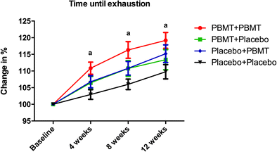
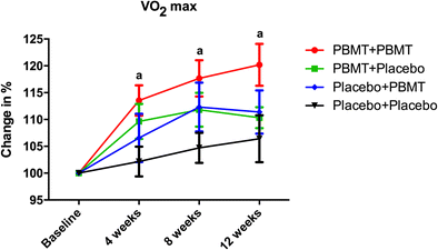
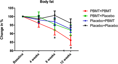
Photobiomodulation therapy (PBMT) employing low-level laser therapy (LLLT) and/or light emitting diode therapy (LEDT) has emerged as an electrophysical intervention that could be associated with aerobic training to enhance beneficial effects of aerobic exercise. However, the best moment to perform irradiation with PBMT in aerobic training has not been elucidated. The aim of this study was to assess the effects of PBMT applied before and/or after each training session and to evaluate outcomes of the endurance-training program associated with PBMT. Seventy-seven healthy volunteers completed the treadmill-training protocol performed for 12 weeks, with 3 sessions per week. PBMT was performed before and/or after each training session (17 sites on each lower limb, using a cluster of 12 diodes: 4 × 905 nm super-pulsed laser diodes, 4 × 875 nm infrared LEDs, and 4 × 640 nm red LEDs, dose of 30 J per site). Volunteers were randomized in four groups according to the treatment they would receive before and after each training session: PBMT before + PBMT after, PBMT before + placebo after, placebo before + PBMT after, and placebo before + placebo after. Assessments were performed before the start of the protocol and after 4, 8, and 12 weeks of training. Primary outcome was time until exhaustion; secondary outcome measures were oxygen uptake and body fat. PBMT applied before and after aerobic exercise training sessions (PBMT before + PBMT after group) significantly increased (p < 0.05) the percentage of change of time until exhaustion and oxygen uptake compared to the group treated with placebo before and after aerobic exercise training sessions (placebo before + placebo after group) at 4th, 8th, and 12th week. PBMT applied before and after aerobic exercise training sessions (PBMT before + PBMT after group) also significantly improved (p < 0.05) the percentage of change of body fat compared to the group treated with placebo before and after aerobic exercise training sessions (placebo before + placebo after group) at 8th and 12th week. PBMT applied before and after sessions of aerobic training during 12 weeks can increase the time-to-exhaustion and oxygen uptake and also decrease the body fat in healthy volunteers when compared to placebo irradiation before and after exercise sessions. Our outcomes show that PBMT applied before and after endurance-training exercise sessions lead to improvement of endurance three times faster than exercise only.

Introduction

Physical activity is recommended and beneficial for both asymptomatic persons and individuals with chronic diseases [1, 2]. Aerobic endurance is considered a useful tool for the assessment of physical fitness and the detection of changes in aerobic fitness resulting from systematic training [3].

Regular aerobic exercise has various beneficial metabolic, vascular, and cardiorespiratory effects [4]. Additionally, it decreases body fat and increases muscle mass, muscle strength, and bone density [5]. Moreover, it improves self-esteem and physical and mental health and reduces the incidence of anxiety and depression [4, 6].

Various ergogenic agents, such as whey protein [7], caffeine [8], creatine [9], and neuromuscular electrical stimulation [10], are currently used to increase the benefits of aerobic training. Photobiomodulation therapy (PBMT) has emerged as an electrophysical intervention that could be associated with aerobic training to enhance beneficial effects of aerobic exercise, since several studies used PBMT to improve physical performance when associated with different kinds of exercise [11, 12, 13, 14].

Several studies have recently used PBMT to improve muscle performance during aerobic activities in healthy adults [15, 16, 17, 18] and postmenopausal women [19, 20]. However, to the best of our knowledge, the best moment to perform irradiation with PBMT in aerobic training has not been yet elucidated.

For instance, the current literature shows that the application of PBMT before progressive aerobic exercise has ergogenic effects and acutely increases the time until exhaustion, covered distance, and pulmonary ventilation and decreases the score of dyspnea during progressive cardiopulmonary test [15]. In addition, PBMT irradiation performed prior to aerobic exercises improves the exercise performance by decreasing the exercise-induced oxidative stress and muscle damage [18] and increasing the oxygen extraction by peripheral muscles [16]. When performed during aerobic training sessions, PBMT improves the quadriceps power and reduces the peripheral fatigue in postmenopausal women [19, 20]. Additionally, when applied after the sessions of endurance-training program, PBMT leads to a greater fatigue reduction than endurance training without PBMT irradiation [17].

Therefore, the optimal moment to perform PBMT in aerobic training is still open to discussion. With this perspective in mind, we aimed to assess the effects of PBMT applied at different time points (before and/or after) of each training session and its potential effects on the outcomes of an endurance-training program (aerobic exercise).

Materials and methods

Study design and protocol

We performed a triple-blind (assessors, therapists, and volunteers), placebo-controlled, randomized clinical trial. The study was conducted in the Laboratory of Phototherapy in Sports and Exercise.

Ethical aspects

All participants signed informed consent prior to enrollment and the study was approved by the research ethics committee of Nove de Julho University (process 553.831) and registered at Clinical Trials.gov (NCT02874976).

Sample

The sample size was calculated assuming a type I error of 0.05 and a type II error of 0.2, based on previous study [21], and the primary established outcome was the time until exhaustion.

Inclusion and exclusion criteria

We recruited 96 healthy volunteers (48 men and 48 women) between 18 and 35 years of age and without training or involvement in a regular exercise program (i.e., exercise more than once per week) [22, 23]. Volunteers were excluded if they had any skeletal muscle injury, used any nutritional supplement or pharmacologic agent, presented with signs or symptoms of any disease (i.e., neurologic, inflammatory, pulmonary, metabolic, oncologic), or had a history of cardiac arrest that might limit performance of high-intensity exercises. Volunteers that were unable to attend a minimum rate of 80% of the training sessions and volunteers with immune diseases that require continuous use of anti-inflammatory drugs were also excluded.

Randomization and blinding procedures

Volunteers were distributed in four experimental groups (24 volunteers in each group) through a simple drawing of lots (A, B, C, or D) that determined the moment they would receive active and/or placebo PBMT treatment:
  • PBMT + PBMT: volunteers were treated with active PBMT before and after each training session.

  • PBMT + placebo: volunteers were treated with active PBMT before and placebo PBMT after each training session.

  • Placebo + PBMT: volunteers were treated with placebo PBMT before and active PBMT after each training session.

  • Placebo + placebo: volunteers were treated with placebo PBMT before and after each training session.

Randomization labels were created by using a randomization table at a central office where a series of sealed, opaque, and numbered envelopes ensured confidentiality. The researcher who programmed the PBMT device (manufactured by Multi Radiance Medical™, Solon, OH, USA) based on the randomization results was not involved in any other procedure of the study. He was instructed not to inform the participants or other researchers of the PBMT program (active or placebo). None of the researchers involved in aerobic endurance-training assessments and data collection knew which program corresponded to active or placebo PBMT.

Identical PBMT devices were used in both programs (active or placebo) by a researcher who was not involved in any phase of the projected data collection to ensure the study blinding. All displays and sounds emitted were identical regardless of the selected program. The active PBMT treatment did not demonstrate discernable amounts of heat [24].

Therefore, volunteers were unable to differentiate between active or placebo treatments. All volunteers were required to wear opaque goggles during treatments to safety and to maintain the triple-blind design.

Procedures

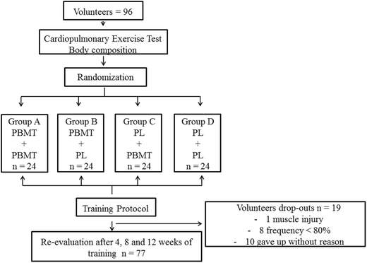
The study included three sessions of aerobic endurance training per week performed over 12 weeks, and each session lasted 30 min; the load for each exercise session (treadmill speed) progressed constantly in order to keep subjects’ heart rate between 70 and 80% from maximum heart rate. The assessments were conducted before the start of the training protocol and after 4, 8, and 12 weeks of training. A summary of the study design is presented in Fig. 1.
Fig. 1

CONSORT flowchart

Cardiopulmonary exercise test

Participants performed a standardized progressive cardiopulmonary exercise test on a treadmill with a fixed inclination of 1% until exhaustion. They began the test with a 3-min warm-up at a velocity of 3 km/h. Next, the treadmill velocity was increased by 1 km/h at 1-min intervals until the velocity of 16 km/h was reached. Participants were instructed to use hand signals to request termination of the test at any time. A 3-min recovery phase at a velocity of 6 km/h was allowed after each test [18]. During testing, we monitored the rates of oxygen uptake (VO2), carbon dioxide production measured with a VO 2000 gas analyzer (Inbrasport, Indústria Brasileira de Equipamentos Médico-Desportivos LTDA, Porto Alegre, RS, Brazil), total time until exhaustion, and heart rate measured with a digital electrocardiograph (Medical Graphs Ergomet, São Paulo, SP, Brazil).

These data were used to evaluate the performance of participants during progressive cardiopulmonary exercise testing, because this test is currently the most widely used in the literature for this purpose [25]. The entire test was monitored by electrocardiogram and blood pressure measurement. If any abnormal heart rate or blood pressure changes were observed or if the test was terminated prematurely on request, the test was stopped, and the volunteer’s data were deleted.

Body composition assessment

Body composition was assessed by the same technician (blinded to volunteer’s allocation in different experimental groups) using the procedures established by ISAK [26]. Measurements of height, body mass, and skinfolds were used to establish the percentage of fat [26].

Aerobic training protocol

Aerobic treadmill training, associated or not with PBMT, was performed three times a week for 12 weeks, each session lasting 30 min, with training intensity kept between 70 and 80% of maximum heart rate [27]; changes in running speed (training load) were constantly performed to achieve the 70–80% heart rate.

Training was interrupted based on the criteria established by the guidelines of the American Heart Association. Training intensity was monitored by a heart rate monitor manufactured by Polar®.

Photobiomodulation therapy

PBMT was applied employing MR4 Laser Therapy Systems outfitted with LaserShower 50 4D emitters (both manufactured by Multi Radiance Medical, Solon, OH, USA). The cluster style emitter contains 12 diodes composing of four super-pulsed laser diodes (905 nm, 0.3125 mW average power, and 12.5 W peak power for each diode), four red LED diodes (640 nm, 15 mW average power for each diode), and four infrared LEDs diodes (875 nm, 17.5 mW average power for each diode).

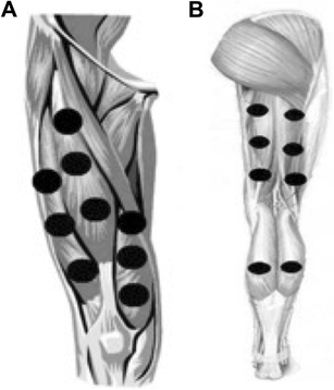
The cluster probe was selected due to the available coverage area and to reduce the number of sites needing treatment. Treatment was applied in direct contact with the skin with a slight applied overpressure to nine sites on extensor muscles of the knee (Fig. 2a), six sites on knee flexors of the knee, and two sites on the calf (Fig. 2b) of both lower limbs [15, 28]. To ensure blinding, the device emitted the same sounds and regardless of the programmed mode (active or placebo). The researcher, who was blinded to randomization and the programming of PBMT device, performed the PBMT.
Fig. 2

a Treatment sites at knee extensor muscles. b Treatment sites at knee flexor and ankle plantar flexor muscles

PBMT parameters and irradiation sites were selected based upon previous positive outcomes demonstrated with the same family of device [13, 15, 28, 29]. Table 1 provides a full description of the PBMT parameters. The volunteers received PBMT or placebo from 5 to 10 min before and/or after aerobic training sessions.


Statistical analysis

The obtained results were tested for their normality through the Shapiro-Wilk test. Since the data showed a normal distribution, two-way ANOVA test with Bonferroni post hoc analysis was applied. The data were described as mean values with the respective standard deviations and both absolute and percentage values were analyzed. Graphical data are described as mean and standard errors of mean (SEM). The level of statistical significance was p < 0.05.

Results

After data collection, we analyzed the results of 77 volunteers of both genders (PBMT + PBMT: 18 volunteers; PBMT + placebo: 21 volunteers; placebo + PBMT: 18 volunteers; and placebo + placebo: 20 volunteers) that had completed the aerobic training protocol after 12 weeks (Fig. 1). None of the recruited volunteers were excluded due abnormal heart rate or blood pressure during the execution of procedures of this study. The characteristics of the volunteers are summarized in Table 2.


As shown in Table 2, no statistically significant differences (p > 0.05) were found for anthropometric variables and baseline data among the different experimental study groups.

Table 3 shows all results of cardiopulmonary progressive test in absolute values for different variables analyzed in all experimental groups of this study. We observed a statistically significant improvement in oxygen uptake when PBMT was performed before and after training sessions (PBMT + PBMT group), comparing baseline values vs 4-, 8-, and 12-week values (p < 0.001). The same was observed for pulmonary ventilation, comparing baseline values vs 8- and 12-week values (p = 0.0018 and p = 0.003, respectively), and for time until exhaustion, comparing baseline values vs 4-, 8-, and 12-week values (p < 0.001).
Table 3

Progressive endurance test variables


Baseline

4 weeks

8 weeks

12 weeks

VO2 (mL/kg/min)

PBMT + PBMT

35.8 ± 9.5

40.2 ± 10.2*

41.5 ± 10.4*

42.5 ± 11.2*

PBMT + Placebo

34.8 ± 7.0

37.6 ± 7.0

38.6 ± 8.0

38.2 ± 7.0

Placebo + PBMT

35.2 ± 8.9

36.6 ± 8.1

38.6 ± 8.3

38.5 ± 8.3

Placebo + placebo

36.2 ± 7.7

36.8 ± 8.0

37.6 ± 7.5

38.4 ± 10.1

VCO2 (mL/kg/min)

PBMT + PBMT

38.7 ± 7.0

40.4 ± 8.6

41.3 ± 7.8

41.4 ± 8.7

PBMT + placebo

38.,5 ± 7.8

39.5 ± 6.6

41.7 ± 7.9

41.9 ± 6.8

Placebo + PBMT

38.5 ± 9.5

38.2 ± 9.5

41.5 ± 8.4

40.7 ± 9.6

Placebo + placebo

38.8 ± 10.6

40.7 ± 9.4

43.1 ± 13.4

40.9 ± 10.5

VE (mL/kg/min)

PBMT + PBMT

73.6 ± 22.8

77.9 ± 21.5

83.5 ± 24.5*

85.3 ± 22.5*

PBMT + Placebo

70.6 ± 20.3

71.0 ± 23.1

78.1 ± 23.0

77.2 ± 22.1

Placebo + PBMT

66.2 ± 25.3

70.6 ± 24.2

73.9 ± 20.6

73.4 ± 20.7

Placebo + placebo

69.9 ± 17.9

70.8 ± 18.8

70.3 ± 22.4

77.1 ± 18.3

Time until exhaustion (s)

PBMT + PBMT

681.5 ± 111.9

752.1 ± 111.7*

787.7 ± 114.2*

808.5 ± 124.5*

PBMT + placebo

698.7 ± 131.1

739.3 ± 142.2

773.4 ± 165.9

792.1 ± 186.9

Placebo + PBMT

693.1 ± 106.9

738.4 ± 116.6

766.1 ± 121.0

797.0 ± 139.0

Placebo + placebo

699.5 ± 137.3

720.2 ± 150.0

741.3 ± 154.3*

766.1 ± 159.8*

Data is expressed in average and standard deviation (±)

VO 2 oxygen uptake, VCO 2 carbon dioxide production, VE pulmonary ventilation

*Statistically significant difference compared to baseline (p < 0.05)

Furthermore, PBMT applied before and after each aerobic exercise training session (PBMT + PBMT group) significantly increased (p < 0.05) the percentage change of oxygen consumption and time-to-exhaustion compared to the group treated with placebo before and after each aerobic exercise training session (placebo + placebo group) from 4th to 12th week. Similarly, PBMT applied before and after each aerobic exercise training session (PBMT + PBMT group) significantly improved (p < 0.05) the percentage change of body fat compared to group treated with placebo before and after each aerobic exercise training session (placebo + placebo group). The outcomes are summarized in Figs. 3, 4, and 5, respectively.

Fig. 3

Percentage of change in time-to-exhaustion. The data are presented in mean and SEM. Letter a indicates statistical significance between PBMT + PBMT and placebo + placebo (p < 0.05)

Fig. 4

Percentage of change in maximum oxygen uptake. The data are presented in mean and SEM. Letter a indicates statistical significance between PBMT + PBMT and placebo + placebo (p < 0.05)

Fig. 5

Percentage of change in body fat. The data are presented in mean and SEM. Letter a indicates statistical significance between PBMT + PBMT and placebo + placebo (p < 0.05)

Discussion

To the best of our knowledge, this is the first study aiming to test the optimal moment to perform PBMT in an aerobic training protocol (before, after, or before and after training). Few studies have assessed chronic effects of PBMT [17, 20, 21]; however, PBMT has been applied at different moments (before, after, or during exercise) of the aerobic training program. Briefly, we observed that the combination of super-pulsed lasers and LEDs applied before and after exercise sessions increased the oxygen uptake, time-to-exhaustion, and reduced body fat in healthy sedentary volunteers after 12 weeks of aerobic training.

Paolillo et al. [20] investigated the effects of PBMT applied during the sessions of aerobic training on the treadmill in 20 postmenopausal women. The training was performed twice a week for 3 months, with an intensity of 85–90% of maximum heart rate. The volunteers received LED therapy with 850 nm, 31 mW/cm2, 30 min irradiation, and 14,400 J applied bilaterally to the tight regions. PBMT increased the exercise tolerance time when compared to the control group. These data corroborate with the results of our study, however, we used different light sources and wavelengths simultaneously (4 × 905 nm super-pulsed lasers, 4 × 875 nm infrared LEDs, and 4 × 640 nm red LEDs) to irradiate the volunteers and we found an increase in exercise tolerance of 13.4%. The magnitude of the difference in outcomes between studies might be related to the used irradiation protocol (in our study, the volunteers were irradiated before and after the aerobic training sessions, while Paolillo et al. [20] irradiated volunteers during the training sessions).

The same authors [21] also investigated the effects of PBMT (infrared LEDs—850 nm) when applied during treadmill training in 45 postmenopausal women. The training was performed twice a week for 6 months, and each training session lasted 45 min. The authors found a significant increase in exercise tolerance, and metabolic equivalents, and a longer duration of Bruce test. In our study, the association of PBMT before and after sessions of the aerobic training program was able to increase the oxygen consumption (with 18.7%) and time-to-exhaustion (with 13.4%) and improve the percentage of change of body fat (with 13.9%) after only 12 weeks of aerobic training.

Duarte et al. [30] evaluated the effects of PBMT (808 nm) associated with aerobic and resistance training performed three times a week for 16 weeks in obese women. The authors found a significant decrease in the percentage of fat and in neck and waist circumference. It is important to highlight that in our study, we observed statistically significant improvement in the percentage of change of body fat (13.9%) after only 12 weeks of aerobic training when associated with PBMT before and after the training sessions. We believe that the association of PBMT before and after training was able to enhance the performance and the tolerance of the volunteers during the aerobic training protocol, favoring the reduction of the body fat at the end of the 12 weeks of training.

It is interesting how outcomes in the fourth week for PBMT + PBMT group were similar to those of placebo + placebo group (or exercise alone) in the 12th week. This means that PBMT with optimal irradiation protocol (before and after exercise training sessions) can increase the endurance capacity of volunteers three times faster than exercise alone.

Regarding the mechanisms of the observed effects, we strongly believe that mitochondrial activity modulation is the key mechanism, despite the fact that our study only focused on clinical and functional aspects and not on mechanisms. Hayworth et al. [31] demonstrated that the activity of cytochrome c oxidase is enhanced by PBMT with a single wavelength in skeletal muscle fibers of rats. More recently, Albuquerque-Pontes et al. [32] showed that PBMT with different wavelengths (660, 830, or 905 nm) was able to increase the expression of cytochrome c oxidase in the intact skeletal muscle tissue in different time windows (5 min to 24 h after irradiation), which means that the muscle metabolism can be improved through the action of PBMT. These findings help us to explain the increase in performance observed by the use of PBMT associated with an aerobic training protocol and provide the rationale for the concurrent use of different wavelengths at the same time, which can represent a therapeutic advantage in various clinical situations.

In fact, different studies have shown that the concurrent use of different light sources and wavelengths enhances muscular performance [13, 14, 15, 28, 29, 33] decreases pain [34


Original Source: https://link-springer-com.colorado.idm.oclc.org/article/10.1007%2Fs10103-017-2396-2

MGH-led study shows light therapy is safe, modulates brain repair, and may benefit patients with moderate traumatic brain injury

- (Publication)
“Light therapy is safe and has measurable effects in the brain. Light therapy could become the first widely-accepted treatment for moderate traumatic brain injury”
View Resource

Light therapy is safe and has measurable effects in the brain, according to a pioneering study by researchers from the Wellman Center for Photomedicine at Massachusetts General Hospital (MGH). Senior investigators Rajiv Gupta, MD, PhD, director of the Ultra-High Resolution Volume CT Lab at MGH and Benjamin Vakoc, PhD, at the Wellman Center led the study, which was supported by a grant from the Department of Defense (DOD) and published in JAMA Network Open September 14th. This study is one of the first, if not the first, prospective, randomized, interventional clinical trials of near-infrared, low-level light therapy (LLLT) in patients who recently suffered a moderate brain injury. If further trials support these findings, light therapy could become the first widely-accepted treatment for this type of injury. TBI is the leading cause of traumatic injury worldwide, and an estimated 69 million people experience such an injury every year. However, there are no treatments for this condition yet, largely because the underlying biological mechanisms are not well understood and it is so challenging to do studies with actual patients in the acute stage of trauma. “The Gulf War put TBI in the headlines,” says Gupta, “because body armor had been greatly improved by then. But there were still brain injuries caused by the shock waves from high powered explosives.” For a variety of reasons, the number of TBIs has increased around the globe since then, but effective treatments are still sorely needed. For this study, a special helmet had to be designed specifically to deliver the therapy, an undertaking that required a mix of medical, engineering and physics expertise. This multidisciplinary team included Gupta, a neuroradiologist, Vakoc, an applied physicist, and others specializing in the development and translation of optical instrumentation to the clinic and biologic laboratories. Both Gupta and Vakoc are also associate professors at Harvard Medical School. “For this study, we designed a practical, near-infrared treatment based on Wellman Center research and working directly with DOD on the vexing problem of TBI, a condition faced by so many,” says Rox Anderson, MD, the center’s director. Another challenge was optimizing the wavelength of the near-infrared LLLT. “Nobody knows how much light you need to get the optimal effect,” explains Lynn Drake, MD, one of the study co-authors and director of business development at the Wellman Center. “We tried to optimize the wavelength, dosing, timing of delivery, and length of exposure.” This was done through a series of pre-clinical experiments led by Anderson. These included multiple preclinical studies led by Michael Hamblin, PhD. Anderson and Hamblin are both co-authors on this paper. Near-infrared LLLT has already been considered for multiple uses, but to date, few if any studies of this technology have been tested and none in patients with TBI. It has been studied in stroke patients and Wellman basic laboratory research suggests it is neuroprotective through a mechanism mediated by specialized intracellular organs called mitochondria. It took several years of research at Wellman to understand the basic mechanism prior to the clinical trial. The randomized clinical trial included 68 patients with moderate traumatic brain injury who were divided into two groups. One group received LLLT, via the special helmet, which delivered the light. Patients in the control group wore the helmet for the same amount of time, but did not receive the treatment. The helmet was designed by Vakoc’s team at Wellman. During the study, the subjects’ brains were tested for neuroreactivity using quantitative magnetic resonance imaging (MRI) metrics and the subjects also underwent neurocognitive function assessment. MRI was performed in the acute (within 72 hours of the injury), early subacute (2-3 weeks), and late subacute (approximately three months) stages of recovery. Clinical assessments were performed during each visit and at six months, using the Rivermead Post-Concussion Questionnaire, with each item assessed on a five-point scale. Twenty-eight patients completed at least one LLLT session and none reported any adverse reactions. In addition, the researchers found that they could measure the effects of transcranial LLLT on the brain. The MRI studies showed statistically significant differences in the integrity of myelin surrounding the neurons of treated patients versus the control group. Both these findings support follow-up trials, especially since there are no other treatments for these patients. The study also showed the light does impact the cells. While it is well established that cells have light receptors, “going into this trial, we had several unanswered questions such as whether the light would go through the scalp and skull, whether the dose was sufficient, and whether it would be enough to engage the neural substrates responsible for repair after TBI,” says Gupta. It’s important to note, he adds, that for this initial study, the researchers focused on patients with moderate traumatic brain injury. That helped to ensure their study could have statistically significant findings because patients in this category are more likely to demonstrate a measurable effect. “It would be much more difficult to see such changes in patients with mild injuries and it is quite likely that in patients with severe brain injuries the effect of light therapy would be confounded by other comorbidities of severe trauma,” says Gupta. He adds that researchers are still very early in the development of this therapy, and it is not known if it could be applied to other types of brain injury, such as chronic traumatic encephalopathy (CTE), which has received a lot of public attention over the last few years. CTE is a progressive degenerative disease associated with a history of repetitive brain trauma such as that experienced by certain types of athletes, most notably football players. This study opens up many possibilities for broader use of photomedicine. “Transcranial LED therapy is a promising area of research, with potential to help various brain disorders where therapies are limited,” says Margaret Naeser, PhD, a prominent researcher in photomedicine and research professor of Neurology at Boston University School of Medicine. She was not affiliated with this particular study. This research was partially supported by grants from Air Force contract FA8650-17-C-9113; Army USAMRAA Joint Warfighter Medical Research Program, contract W81XWH-15-C-0052; and Congressionally Directed Medical Research Program W81XWH-13-2-0067. About the Massachusetts General Hospital Massachusetts General Hospital, founded in 1811, is the original and largest teaching hospital of Harvard Medical School. The MGH Research Institute conducts the largest hospital-based research program in the nation, with an annual research budget of more than $1 billion and comprises more than 8,500 researchers working across more than 30 institutes, centers and departments. In August 2020 the MGH was named #6 in the nation by U.S. News & World Report in its list of "America’s Best Hospitals."


Original Source: https://www.massgeneral.org/news/press-release/Mgh-led-study-shows-light-therapy-is-safe-modulates-brain-repair-and-may-benefit-patients-with-moderate-traumatic-brain-injury

Light-emitting diode therapy in exercise-trained mice increases muscle performance, cytochrome c oxidase activity, ATP and cell proliferation

Cleber Ferraresi, Nivaldo Antonio Parizotto, Marcelo Victor Pires de Sousa, Beatriz Kaippert, Ying?Ying Huang, Tomoharu Koiso, Vanderlei Salvador Bagnato, Michael R. Hamblin - Wiley Online Library/ 09-01-2015 (Publication)
This research showed that the light group had significantly more ATP concentration than the control group.
View Resource

Abstract

Light-emitting diode therapy (LEDT) applied over the leg, gluteus and lower-back muscles of mice using a LED cluster (630 nm and 850 nm, 80 mW/cm2, 7.2 J/cm2) increased muscle performance (repetitive climbing of a ladder carrying a water-filled tube attached to the tail), ATP and mitochondrial metabolism; oxidative stress and proliferative myocyte markers in mice subjected to acute and progressive strength training. Six bi-daily training sessions LEDT-After and LEDT-Before-After regimens more than doubled muscle performance and increased ATP more than tenfold. The effectiveness of LEDT on improving muscle performance and recovery suggest applicability for high performance sports and in training programs.


Positioning of the mice and light-emitting diode therapy (LEDT) applied on mouse legs, gluteus and lower-back muscles without contact.

Introduction

Low-level laser (light) therapy has several applications in medicine such as treatment of pain 1, 2, tendinopathies 3 and acceleration of tissue repair 2, 4. Since the 1960s when the first laser (Light Amplification by Stimulated Emission of Radiation) devices were constructed, many applications of this therapy and its mechanisms of action have been investigated around the world 5.

Light therapy can be delivered by different light sources such as diode lasers or light emitting diodes (LEDs). These light sources differ in monochromaticity and coherence, since diode lasers are coherent with a tiny spectral bandwidth and less divergence of the light beams compared to the light emitted by LEDs 5. The spectral regions generally used for light therapy range between red (600 nm) to near infrared (1,000 nm) with total power in range of 1 mW–500 mW and power density (irradiance) in the range of range 1 mW–5 W/cm2 5. These lasers and LEDs are considered to produce equivalent effects on the tissue if the dose of light delivered/applied is in accordance with the possible biphasic dose?response previously reported 5-7. The light?tissue interaction depends on light absorption by specific structures in the cells that are known as chromophores 8-11.

Recently light therapy using lasers and LEDs has been used to increase muscle performance in exercises involving strength 12 or fatigue resistance 13-15; and light therapy may have a role to play in preparing athletes competing in high performance sports. Recent reviews have reported positive effects of light therapy on muscle performance, highlighting protection from exercise?induced muscle damage 16; an increased number of repetitions in maximum exertion tests 17; increased workload, torque and muscle fatigue resistance in training programs; as well as an overview of the main possible mechanisms of action of the light therapy on muscle tissue 18.

Several biological factors govern success or optimum performance in sports that involve high?intensity exercise, or alternatively involve endurance exercise, that both require muscle adaptation during pre?competition training programs. Among these factors are the depletion of the energy supply for muscle contraction which comprises adenosine triphosphate (ATP) and glycogen; accumulation of possibly deleterious metabolites from energy metabolism such as lactate, adenosine diphosphate (ADP), adenosine monophosphate (AMP), ions Ca2+ and H+; production of reactive oxygen species (ROS) 19-22; and the recovery process from microlesions or muscle damage 23. Light therapy seems to be able to benefit all these ”limitations” since its mechanism of action involves the improvement of mitochondrial metabolism and increased ATP synthesis 24, 25 owing to increased activity of cytochrome c oxidase (COX) in the electron transport chain (ETC) 9, 25, 26; reduction of reactive oxygen species (ROS) or improvement of oxidative stress defense 27, 28; and can stimulate faster muscle repair due to an increased proliferation and differentiation of muscle cells 29.

Experimental and clinical trials with different methodologies have reported the benefits of light therapy on muscle performance when applied before 15, 30, 31 or after exercise 12, 13, 32. However there is no consensus about the best time regimen for use of light therapy 18. The best wavelength (red or infrared) to stimulate muscle cells and increase muscle performance is also unclear.

In the current study we used an experimental model of mice exercising on a ladder similar to that reported in a previous study 33, in order to simulate a clinical strength training program that would allow us to identify which light therapy regimen would be better to increase muscle performance. Four different regimens of light therapy were applied to the mouse leg, gluteus and lower?back muscles during a training program: sham; before; before?after; and after each training session. Light therapy was delivered from LEDs (LEDT) with two simultaneous wavelengths (red and infrared). Assessment of muscle performance (load, number of repetitions, muscle work and power), markers of cellular energy and metabolism (ATP, glycogen and COX), oxidative stress markers (protein carbonyls, glutathione, catalase activity, lipid peroxidation, protein thiols) and muscle cell proliferation (BrdU – 5?bromo?2′?deoxyuridine) and adult myonuclei (DAPI – 4′,6?diamidino?2?phenylindole) were carried out.

Materials and methods

Animals

This study was performed with 8 week?old male Balb/c mice, weighing on average 22.22 g (SEM 0.24), housed at five mice per cage and kept on a 12 hour light 12 hour dark cycle. The 22 animals were provided by Charles River Inc and were provided with water and fed ad libitum at the animal facility of Massachusetts General Hospital. All procedures were approved by the IACUC of Massachusetts General Hospital (protocol #2014N000055) and met the guidelines of the National Institutes of Health.

Experimental groups

Twenty?two animals were randomly allocated into 4 exercise groups with 5 animals in each group, and 2 animals were allocated into an ”absolute” control group:

Ladder

An inclined ladder (80°) with dimensions of 100 cm × 9 cm (length and width, respectively) with bars spaced at 0.5 cm intervals was used in this study as reported in a previous study 33 (Figure 1).

Figure 1

Open in figure viewerPowerPoint

Ladder. Inclined ladder (80°) with 100 cm × 9 cm (length and width, respectively) used for the training program and muscle performance assessments. Falcon tube filled with water and attached to the mouse tail.

Load

A Falcon tube (50 ml) was filled with measured volumes of water and weighed using a precise scale. The target load was achieved adding or removing water from the tube and then this tube was attached to the mouse tail using adhesive tape (Figure 1). All loads were calculated in grams.

Procedures

The schedule of the various exercise procedures is described in Table 1.

Table 1. Schedule for exercise procedures

Day

Procedure

# repetitions

Load

Day 1

Familiarization

4 × 10 = 40

zero

Day 2

3RM baseline

3

Starting at 2 × BWa

Day 3

Training 1

5 × 10 = 50

0.8 × 3RMb

Day 5

Training 2

5 × 10 = 50

0.9 × 3RM

Day 7

Training 3

5 × 10 = 50

1.0 × 3RM

Day 9

Training 4

5 × 10 = 50

1.1 × 3RM

Day 11

Training 5

5 × 10 = 50

1.2 × 3RM

Day 13

Training 6

5 × 10 = 50

1.3 × 3RM

Day 14

3RM final

3

Starting at 3 × BW

Familiarization with ladder?climbing

All experimental groups, except Control group, were familiarized with climbing the ladder one day before the start of muscle performance assessment and training. The familiarization procedure was 4 sets of 10 climbs on the ladder (repetitions) with rest periods of 2 minutes between individual sets. No load was attached to the mouse tail during this procedure.

Three repetitions maximum load (3RM)

This test was the first evaluation of muscle performance and was set as the average of the maximum load carried by each animal during 3 consecutive full climbs of the inclined ladder (3RM). Slight pressure with tweezers was applied on mouse tail if the animal stopped during a climb. The test was stopped when mice were not able to climb or lost their grip on the ladder due to failure of concentric muscle contraction. The first attempt included a load corresponding to 200% of the individual mouse body weight. A maximum of 3 climb attempts was applied. If a mouse finished the climb the load was increased by 10% for the next climb, while if the mouse failed to finish a climb, the load was decreased by 10% for the next climb. The 3RM evaluation was performed twice; the first time was 24 h after familiarization procedure (baseline) and the second time was 24 h after the last training session (final).

Acute strength training protocol

After 24 h from initial 3RM baseline assessment, all experimental groups, except Control, were subjected to 6 training sessions carried out on alternate days (every 48 h). Each training session consisted of 5 sets of 10 repetitions (climbs) on the ladder with a rest period of 2 minutes between each set. If the animal could not complete a set or failed during a climb, the distance climbed (in cm) was measured and the rest period was started immediately. During some repetitions, a slight pressure on the mouse tail was performed with tweezers to stimulate the animal to climb and complete the exercise. If after three applications of gentle pressures the mouse could not resume climbing, and stopped or lost its grip on the ladder, the set of repetitions was stopped and the rest interval was started.

The number of repetitions in each set was measured as well as the time spent to complete the exercise. These data were used to calculate the muscle work and muscle power in each training session. The load of each training session was progressively increased and calculated as percentages of the 3RM (in grams) measured at baseline as follows: first training (80%), second training (90%), third training (100%), fourth training (110%), fifth training (120%) and sixth training (130%).

Light?emitting diode therapy (LEDT)

A non?commercial cluster of 40 LEDs (20 red – 630 ± 10 nm; 20 infrared – 850 ± 20 nm) with diameter of 76 mm was used in this study. A complete description of the LEDT parameters is presented in Table 2. The optical power reaching the surface of the mouse skin was measured with an optical energy meter PM100D Thorlabs® fitted with a sensor S142C (area of 1.13 cm2). All mice (except mice in Control) were shaved and fixed on a plastic plate using adhesive tapes. Afterwards, in accordance with experimental group, these animals were treated with LEDT over both legs, gluteus and lower?back muscles at a distance of 45 mm (without contact) (Figure 2). Irradiation lasted 90 s per session with fixed parameters as described in Table 1. LEDT placebo had no energy (0 J) and no power (0 mW) applied over the targeted muscles. The light dose was based on the possible biphasic dose response reported previously 5, 6. Moreover, dual wavelengths were chosen to function at the same time in this study based on specificities of the chromophores in the cells and therefore optimizing the effects of the light therapy (LEDT) by a double band of absorption 8-11.

Figure 2

Open in figure viewerPowerPoint

LEDT. Positioning of the mice and light?emitting diode therapy (LEDT) applied on mouse legs, gluteus and lower?back muscles without contact.


Muscle performance

The 3RM test was the first evaluation for muscle performance. This test measured the maximum load (in grams) carried by each animal during 3 consecutive full climbs on the inclined ladder.

During each training session the load, number of repetitions (rep), distance climbed and time spent to complete each repetition were recorded. These data were used to calculate muscle work and power.

Although the ladder had a total length of 100 cm available the maximum distance available to climb was set at 70 cm in order to avoid the load touching the floor. Thereby the muscle work was calculated as follows:

Work (J) = mgh

where ”m” is mass of the load (grams converted to kilogram) in each training session plus mouse body mass (values converted to kilogram); ”g” is acceleration due to gravity and ”h” is the distance climbed (converted to meters). Results were obtained in Joules (J) and presented as average ± standard error of mean (SEM) for each group at each training session.

Muscle power was calculated from results of muscle work (J) and time spent (s) to perform all repetitions of each set at all training sessions as follows:

Power (mW) = J/s

where ”J” is Joule and represents the muscle work performed and ”s” is time in seconds. Result were obtained in milliwatts (mW) and presented as average ± standard error of mean (SEM) per each group at each training session.

Muscular ATP

The gastrocnemius muscle from one leg of each animal was used for analysis of muscular ATP. Muscle samples were thawed in ice for 5 min, homogenized at a proportion of 3–4 mg of tissue to 500 µl of 10% perchloric acid (HClO4) following procedures previously published 34. Afterwards, an aliquot of 10 µl of the muscle homogenate plus 40 µl of CellTiter Glo Luminescent Cell Viability Assay mix (Promega), totaling 50 µl, were placed in the well microplate (CostarTM 96?Well White Clear?Bottom Plates). Luminescence signals were measured in a SpectraMax M5 Multi?Mode Microplate Reader (Molecular Devices, Sunnyvale, CA) with integration time of 5 s to increase low signals 34. A standard curve was prepared using ATP standard (Sigma) according to manufacturer's guidelines and then ATP concentration was calculated in nanomol (nmol) per milligram (mg) of protein. An aliquot of muscle homogenate was used to quantify the total protein by QuantiProTM BCA Assay kit (Sigma?Aldrich) following manufacturer's guidelines.

Muscular glycogen

Quadriceps femoris muscles were thawed in ice for 30 min and muscular glycogen was measured in 50 mg of quadriceps femoris tissue homogenized with 6 N NaOH at a proportion of 50 mg/ml. A standard curve was prepared using absolute ethanol (100%), K2SO4 (10%), phenol (4.1%) and 1 mM of glucose (2%) according to Dubois et al. 35. Optical density was read at 480 nm in spectrophotometer (EvolutionTM 300 UV?Vis, software VISPRO – Thermo Scientific). Data were normalized per mg of muscle tissue.

Oxidative stress markers

Protein carbonyl: Quadriceps femoris muscles were homogenized in deionized water (dH2O) at a proportion of 10 mg/200 µl. Protein carbonyl content was quantified using Protein Carbonyl Content Assay kit (Biovision) with the colorimetric method and following manufacturer's guidelines. All results were normalized per total protein quantified by QuantiProTM BCA Assay kit (Sigma?Aldrich) following manufacturer's guidelines.

Glutathione: Quadriceps femoris muscles were homogenized in 100 mM ice cold phosphate buffer (pH = 7.4) at a proportion of 10 mg/250 µl. Phosphate buffer was prepared with dibasic (Na2HPO4) and monobasic (NaH2PO4) sodium phosphate at equal proportions. Total and oxidized glutathione analysis was carried out with Glutathione Colorimetric Assay kit (ARBOR Assays) following manufacturer's guidelines. In addition, all results were normalized per total protein of the samples using QuantiProTM BCA Assay kit (Sigma?Aldrich) following manufacturer's guidelines.

Catalase activity: Quadriceps femoris muscles were homogenized in cold assay buffer provided in a Catalase Activity Assay kit (Biovision) at a proportion of 50 mg/100 µl. This analysis used the colorimetric method and followed manufacture's guidelines.

Lipid peroxidation using TBARS (Thiobarbituric Acid Reactive Substances): Quadriceps femoris muscles were homogenized with RIPA Buffer (Sigma?Aldrich) at a proportion of 25 mg/250 µl. Next, TBARS Colorimetric Assay kit (Cayman Chemical) was used following manufacturer's guidelines.

Protein Thiols: Quadriceps femoris muscles were homogenized in ice cold 100 mM phosphate buffer at a proportion of 10 mg/250 µl. Next, a Fluorescent Protein Thiol Detectiont kit (ARBOR Assays) was used following manufacturer's guidelines. In addition, all results were normalized per total protein quantified by QuantiProTM BCA Assay kit (Sigma?Aldrich) following manufacturer's guidelines.

Immunofluorescence analyses

5?bromo?2′?deoxyuridine (BrdU): BrdU reagent (Sigma?Aldrich) was diluted in saline solution (PBS) at a concentration of 10 mg/ml. Next, during the last 8 days of the experiment all animals (including Control group) received a single daily intra peritoneal injection (50 mg/kg) of BrdU. Mice were anesthetized and submitted to surgical procedures described previously. Gastrocnemius muscles were embedded in paraffin, cut in axial slices of 5 µm thickness from the muscle belly region by a microtome and mounted on slides for immunohistochemical procedures. Briefly, slides were deparaffinized with graded ethanol and then passed through antigen retrieval solution in a water bath pre?heated at 98 °C for 30 min. Afterwards slides were washed and incubated for 15 min at room temperature with 0.1% Triton X?100 TBS for cell membrane permeabilization, washed again and incubated for 30 min in protein blocking solution consisting of 3% BSA (Bovine Serum Albumin – Sigma) and 10% goat serum in TBS. Next, slides were immunostained with sheep anti?BrdU (Ab1893 – Abcam, Cambridge, MA) at 1 : 50 working concentration and selected anti?sheep (Alexa Fluor® 647 – Invitrogen) fluorescent secondary antibody matched to the primary antibody to stain at 1 : 200 working concentration. Finally, slides were cover?slipped with mounting media containing DAPI (4′,6?diamidino?2?phenylindole) (Invitrogen). Cells positively stained for BrdU were imaged using confocal microscope (Olympus America Inc. Center Valley, PA, USA) from three random fields. BrdU and DAPI staining were quantified using software Image J (NIH, Bethesda, MD).

Cytochrome c oxidase subunit IV (COX IV): Gastrocnemius muscles were subjected to the same procedures described for BrdU staining. Slides were immunostained with rabbit anti?COX IV (Cell Signaling Technology®) at 1 : 500 working concentration and selected anti?rabbit (Alexa Fluor® 680 – Invitrogen) secondary antibody matched with primary antibody to stain at 1 : 200 working concentration. Cells positively stained for COX IV were imaged using confocal microscopy as above and then the red channel of the exported images was changed to yellow.

Statistical analysis

Shapiro?Wilk's W test verified the normal distribution of the data. All experimental groups subjected to training protocols were compared at each training session for number of repetitions, muscle work and muscle power using one?way analysis of variance (ANOVA) and Tukey HSD post?hoc test. The load of 3RM among these same groups was compared by Two?way ANOVA with repeated measures (baseline versus final) and Tukey HSD post?hoc test. For muscular ATP, glycogen, oxidative stress markers and immunofluorescence stains, all experimental groups were compared by one?way ANOVA and Tukey's HSD post?hoc test. Significance was set at p < 0.05.


Results

Muscle performance

3RM: The final load 3RM was significantly higher (p < 0.05) in all experimental groups at the end of the experiment period compared to baseline. The final load of LEDT?After (92.28 g, SEM 0.82) was higher than LEDT?Sham (59.58 g, SEM 5.28; p < 0.001) and LEDT?Before (78.98 g, SEM 1.96; p = 0.020). In addition, LEDT?Sham had a significantly lower final load (p < 0.001) compared to LEDT?Before as well as LEDT?Before/After (83.91 g, SEM 1.49) (Figure 4A).

Figure 4

Open in figure viewerPowerPoint

Muscle performance (n = 5 animals per group). (A) Baseline and Final test of 3 repetitions maximum (3RM) measuring the total load carried by mice during this test. * statistical significance (p < 0.05) comparing the final 3RM load between groups. (B) Number of repetitions or climbs performed by each group treated with different regimens of LEDT during the progressive training program. (C) Muscle power developed by each group treated with different regimens of LEDT during the progressive training program. (D) Muscle work developed by each group treated with different regimens of LEDT during the progressive training program. * statistical significance (p < 0.05) compared to LEDT?Sham. # statistical significance (p < 0.05) compared to LEDT?After. & statistical significance (p < 0.05) compared to LEDT?Before. Abbreviations: LEDT = light?emitting diode therapy; LEDT?Sham (Sham – S) = LEDT placebo (LEDT device in placebo mode) on muscles immediately before (5 minutes) each training session on ladder; LEDT?Before (Before – B) = LEDT applied on muscles immediately before (5 minutes) each training session on ladder; LEDT?Before?After (Before?After – A?B) = LEDT applied on muscles immediately before (5 minutes) and immediately after (5 minutes) each training session on ladder; LEDT?After (After – A) = LEDT applied on muscles immediately after (5 minutes) each training session on ladder. The load of 3RM at baseline versus final was analyzed by Two?way analysis of variance (ANOVA) with repeated measures. Number of repetitions, muscle work and power were analyzed by One?way ANOVA.

Number of repetitions: There were significantly differences (p < 0.05) between all groups in each training session (Figure 4B). At 80% of 3RM (first session): animals in LEDT?Before and LEDT?Before?After groups performed more repetitions compared to animals in LEDT?Sham and LEDT?After (p < 0.01) groups. At 90% of 3RM (second session): animals in LED?Sham group performed fewer repetitions than animals in LEDT?Before, LEDT?Before?After and LEDT?After groups (p < 0.001). At 100% of 3RM (third session): animals in LEDT?Sham group performed fewer repetitions compared to animals in LEDT?Before (p = 0.014), LED?Before?After (p = 0.010) and LEDT?After (p = 0.002) groups. At 110% of 3RM (fourth session): animals in LEDT?Sham group performed fewer repetitions than animals in LEDT?Before?After (p = 0.013) and LEDT?After (p = 0.009) groups. At 120% of 3RM (fifth session): animals in LEDT?After group performed more repetitions than animals in LEDT?Before (p = 0.022) and LEDT?Sham (p < 0.001) groups. In addition, animals in LEDT?Sham performed fewer repetitions than animals in LEDT?Before (p = 0.022), LEDT?Before?After and LEDT?After (p < 0.001) groups. At 130% of 3RM (sixth session): animals in LEDT?Before?After and LEDT?After groups performed more repetitions than animals in LEDT?Sham (p < 0.001) and LEDT?Before (p < 0.01) groups.

Muscle Power: At 80% of 3RM there were no significant differences among all groups (p > 0.05). At 90% of 3RM: animals in LEDT?Sham group had lower muscle power compared to animals in LEDT?Before, LEDT?Before?After and LEDT?After (p < 0.01) groups. At 100% of 3RM: animals in LEDT?Sham group had lower muscle power than animals in LEDT?Before?After (p = 0.025) and LEDT?After (p = 0.007) groups. At 110% of 3RM: animals in LEDT?Before?After group developed more muscle power than animals in LEDT?Sham (p < 0.001) and LEDT?Before (p = 0.013) groups. In addition, animals in LEDT?After group had more muscle power than animals in LEDT?Sham (p = 0.002) group. At 120% of 3RM: animals in LEDT?Before?After and LEDT?After groups developed more muscle power than animals in LEDT?Sham and LEDT?Before (p < 0.001) groups. At 130% of 3RM: animals in LEDT?Before?After group developed more muscle power than animals in LEDT?Sham and LEDT?Before (p < 0.001) as well as LEDT?After (p = 0.001) groups. In addition, animals in LEDT?After group had more muscle power than animals in LEDT?Sham (p < 0.001) and LEDT?Before (p = 0.004) groups. Finally, animals in LEDT?Before group had major muscle power than animals in LEDT?Sham (p = 0.020) group (Figure 4C).

Muscle Work: Similar to results presented in Figure 4B, at 80% of 3RM only animals in LEDT?Before and LEDT?Before?After groups performed more muscle work compared to LEDT?Sham (p < 0.05) group (Figure 4D). At 90% of 3RM: animals in LEDT?Sham group performed less muscle work than animals in LEDT?Before, LEDT?Before?After and LEDT?After (p < 0.001) groups. These results were similar at 100% of 3RM (p < 0.001). At 110% of 3RM: animals in LEDT?Sham group had lower muscle work compared to animals in LEDT?Before?After (p = 0.015) and LEDT?After (p = 0.011) groups. At 120% of 3RM: animals in LEDT?Sham group performed lower muscle work compared to animals in LEDT?Before (p = 0.027) and LEDT?Before?After and LEDT?After (p < 0.001) groups. In addition, animals in LEDT?After group performed more muscle work than animals in LEDT?Before (p = 0.026) group. At 130% of 3RM: animals in LEDT?Before?After and LEDT?After groups performed more muscle work than animals in LEDT?Sham (p < 0.001) and LEDT?Before (p < 0.01) groups (Figure 4D).

Muscle ATP content

Animals in LEDT?After group had significantly (p < 0.001) more ATP concentration (1,367.64 nmol/ mg protein, SEM 105.30) compared to animals in LEDT?Sham (15.85 nmol/mg protein, SEM 5.14), LEDT?Before (81.00 nmol/ mg protein, SEM 10.11), LEDT?Before?After (687.62 nmol/ mg protein, SEM 11.76) and Control (17.53 nmol/mg protein, SEM 7.47) groups. In addition, animals in LEDT?Before?After group had also major contents of ATP compared to animals in LEDT?Before, LEDT?Sham and Control (p < 0.001) groups (Figure 5A).


Figure 5

Open in figure viewerPowerPoint

Muscular ATP and glycogen contents (n = 5 animals per group). (A) Adenosine triphosphate (ATP) contents in gastrocnemius muscle after the training program. (B) Glycogen contents in quadriceps femoris muscles after the training program. * statistical significance (p < 0.05). Abbreviations: LEDT = light?emitting diode therapy; LEDT?Sham (Sham – S) = LEDT placebo (LEDT device in placebo mode) on muscles immediately before (5 minutes) each training session on ladder; LEDT?Before (Before – B) = LEDT applied on muscles immediately before (5 minutes) each training session on ladder; LEDT?Before?After (Before?After – A?B) = LEDT applied on muscles immediately before (5 minutes) and immediately after (5 minutes) each training session on ladder; LEDT?After (After – A) = LEDT applied on muscles immediately after (5 minutes) each training session on ladder. Control (C) = no exercise or muscle performance assessment. Comparisons among all groups were conducted using One?way analysis of variance (ANOVA).


Muscle glycogen content

Animals in LEDT?After (137.76 nmol/mg tissue, SEM 11.40) and LEDT?Before?After (144.44 nmol/ mg tissue, SEM 16.23) groups had significantly higher concentrations of glycogen in quadriceps femoris muscles (p < 0.001) compared to animals in LEDT?Sham (31.36 nmol/mg tissue, SEM 7.45), LEDT?Before (52.76 nmol/mg tissue, SEM 6.53) and Control (58.78 nmol/ mg tissue, SEM 7.17) groups (Figure 5B).

Oxidative stress markers

Total glutathione: Animals in Control group (1.33 µM/µg protein, SEM 0.11) had a significantly higher concentration of total glutathione compared to animals in LEDT?Sham (0.097 µM/µg protein, SEM 0.046; p = 0.005) and LEDT?Before (1.00 µM/µg protein, SEM 0.02; p = 0.010) groups (Figure 6A).

Figure 6

Open in figure viewerPowerPoint

Oxidative stress markers (n = 5 animals per group) in quadriceps femoris muscles. (A) Total Glutathione (reduced glutathione – GSH). (B) Oxidized Glutathione (GSSG). (C) Protein Carbonyl. (D) Catalase activity. (E) Lipid peroxidation using TBARS (Thiobarbituric Acid Reactive Substances). (F) Protein Thiol. * statistical significance (p < 0.05). Abbreviations: LEDT = light?emitting diode therapy; LEDT?Sham (Sham – S) = LEDT placebo (LEDT device in placebo mode) on muscles immediately before (5 minutes) each training session on ladder; LEDT?Before (Before – B) = LEDT applied on muscles immediately before (5 minutes) each training session on ladder; LEDT?Before?After (Before?After – A?B) = LEDT applied on muscles immediately before (5 minutes) and immediately after (5 minutes) each training session on ladder; LEDT?After (After – A) = LEDT applied on muscles immediately after (5 minutes) each training session on ladder. Control (C) = no exercise or muscle performance assessment. Comparisons among all groups were conducted using One?way analysis of variance (ANOVA).

Oxidized glutathione: Animals in LEDT?Sham group (0.005 µM/µg protein, SEM 0.001) had significantly minor concentration of glutathione oxidized compared to animals in LEDT?Before (0.20 µM/µg protein, SEM 0.002; p = 0.015), LEDT?Before?After (0.035 µM/µg protein, SEM 0.003; p < 0.001), LEDT?After (0.041 µM/µg protein, SEM 0.003; p < 0.001) and Control (0.027 µM/µg protein, SEM 0.007; p = 0.006) groups. In addition, animals in LEDT?Before group had significantly minor concentration of oxidized glutathione compared to animals in LEDT?After (p < 0.001) and LEDT?Before?After (p = 0.024) groups (Figure 6B).

Protein carbonyl: Animals in LEDT?After group (1.40 nmol/µg protein, SEM 0.15) had significantly lower concentrations of protein carbonyls compared to animals in LEDT?Sham (6.31 nmol/µg protein, SEM 1.09; p = 0.030), LEDT?Before (6.81 nmol/µg protein, SEM 1.21; p = 0.040) and LEDT?Before?After (8.27 nmol/µg protein, SEM 2.35; p = 0.008) groups (Figure 6C).

Catalase activity: Animals in LEDT?Sham group (2.11 nmol/min/ml, SEM 0.10) had significantly lower catalase activity (p < 0.01) compared to animals in LEDT?Before?After (4.33 nmol/min/ml, SEM 0.62), LEDT?After (4.22 nmol/min/ml, SEM 0.37) and Control (4.47 nmol/min/ml, SEM 0.52) groups (Figure 6D).

Lipid peroxidation using TBARS: There were no significant differences between any of the groups (p > 0.05) assessed. Animals in Control group had a concentration of 21.29 µM (SEM 1.13); animals in LEDT?Sham had 21.12 µM (SEM 2.86); animals in LEDT?Before had 23.87 µM (SEM 1.13); animals in LEDT?Before?After had 19.19 µM (SEM 1.01) and animals in LEDT?After had 19.55 µM (SEM 1.24) (Figure 6E).

Protein Thiols: There were no sig


Original Source: https://onlinelibrary-wiley-com.colorado.idm.oclc.org/doi/full/10.1002/jbio.201400087

Biphasic Dose Response in Low Level Light Therapy – An Update

Ying-Ying Huang, Sulbha K Sharma, Michael R Hamblin - Published online 2011 Sep 2. doi: 10.2203/dose-response.11-009.Hamblin (Publication)
This research talks about the controversial bi-phasic response from light and laser therapy.
View Resource

Low-level laser (light) therapy (LLLT) has been known since 1967 but still remains controversial due to incomplete understanding of the basic mechanisms and the selection of inappropriate dosimetric parameters that led to negative studies. The biphasic dose-response or Arndt-Schulz curve in LLLT has been shown both in vitro studies and in animal experiments. This review will provide an update to our previous (Huang et al. 2009) coverage of this topic. In vitro mediators of LLLT such as adenosine triphosphate (ATP) and mitochondrial membrane potential show biphasic patterns, while others such as mitochondrial reactive oxygen species show a triphasic dose-response with two distinct peaks. The Janus nature of reactive oxygen species (ROS) that may act as a beneficial signaling molecule at low concentrations and a harmful cytotoxic agent at high concentrations, may partly explain the observed responses in vivo. Transcranial LLLT for traumatic brain injury (TBI) in mice shows a distinct biphasic pattern with peaks in beneficial neurological effects observed when the number of treatments is varied, and when the energy density of an individual treatment is varied. Further understanding of the extent to which biphasic dose responses apply in LLLT will be necessary to optimize clinical treatments.

Keywords: low level laser therapy, photobiomodulation, biphasic dose response, reactive oxygen species, nitric oxide, traumatic brain injury

Go to:

INTRODUCTION

Low level laser (light) therapy (LLLT) employs visible (generally red) or near-infrared light generated from a laser or light emitting diode (LED) system to treat diverse injuries or pathologies in humans or animals. The light is typically of narrow spectral width between 600nm – 1000nm. The fluence (energy density) used is generally between 1 and 20 J/cm2 while the irradiance (power density) can vary widely depending on the actual light source and spot size; values from 5 to 50 mW/cm2 are common for stimulation and healing, while much higher irradiances (up to W/cm2) can be used for nerve inhibition and pain relief. LLLT is typically used to promote tissue regeneration, reduce swelling and inflammation and relieve pain and is often applied to the injury for 30 seconds to a few minutes or so, a few times a week for several weeks. Unlike other medical laser procedures, LLLT is not an ablative or thermal mechanism, but rather a photochemical effect comparable to photosynthesis in plants whereby the light is absorbed and exerts a chemical change.

Within a decade of the introduction of LLLT in the 1970s it was realized that more does not necessarily mean better. The demonstration of the biphasic dose response curve in LLLT has been hampered by disagreement about exactly what constitutes a “dose”. Many practitioners concentrate on fluence as the principle metric of dose, while others prefer irradiance or illumination time. The use of very small spot sizes by some practitioners has led to the assertion that they delivered hundreds of mW/cm2 from a 50 mW laser. While this statement is mathematically correct it can give the impression that much higher doses of light were given than actually were delivered.

Two years ago we reviewed (Huang et al. 2009) the biphasic dose response in LLLT and found many reports in the literature concerning biphasic dose responses observed in cell cultures, some in animal experiments but no clinical reports. We now believe that the time is right to revisit this interesting topic for two reasons. Firstly because we have found more instances in our laboratory both in vitro with cultured cortical neurons, and in vivo with LLLT of traumatic brain injuries in mouse models. Secondly because advances have been made in mechanistic understanding of how LLLT works at a cellular level that may explain why a little light may be beneficial and at the same time a lot of light might be harmful.

Go to:

MECHANISMS OF LOW LEVEL LIGHT THERAPY

Basic photobiophysics and photochemistry

According to the First Law of Photochemistry, the photons of light must be absorbed by some molecular photoacceptors or chromophores for photochemistry to occur (Sutherland 2002).The mechanism of LLLT at the cellular level has been attributed to the absorption of monochromatic visible and near infrared (NIR) radiation by components of the cellular respiratory chain (Karu 1989). Phototherapy is characterized by its ability to induce photobiological processes in cells. The effective tissue penetration of light and the specific wavelength of light absorbed by photoacceptors are two of the major parameters to be considered in light therapy. In tissue there is an “optical window” that runs approximately from 650 nm to 1200 nm where the effective tissue penetration of light is maximized. Therefore the use of LLLT in animals and patients almost exclusively involves red and near-infrared light (600–1100-nm) (Karu and Afanas’eva 1995). The action spectrum (a plot of biological effect against wavelength) shows which specific wavelengths of light are most effectively used for biological endpoints as well as for further investigations into cellular mechanisms of phototherapy (Karu and Kolyakov 2005). Fluence (J/cm2) is often referred to as “dose”, though many authors and practitioners of LLLT also refer to energy (Joules) as dose. Not only is this confusing to the novice student of LLLT but it also assumes that the product of power and time (and more importantly power density and time) is the goal rather than the right combination of individual values. This lack of reciprocity has been shown many times before and since our first paper on biphasic dose response and several more authors have reported finding these effects since. Examples of recently published “dose-rate” effects are also reviewed later in this article.

Mitochondrial Respiration and Cytochrome c oxidase

Mitochondria play an important role in energy generation and metabolism and are involved in current research about the mechanism of LLLT effects. The absorption of monochromatic visible and NIR radiation by components of the cellular respiratory chain has been considered as the primary mechanism of LLLT at the cellular level (Karu 1989). Cytochrome c oxidase (Cco) is proposed to be the primary photoacceptor for the red-NIR light range in mammalian cells. Absorption spectra obtained for biological responses to light were found to be very similar to the absorption spectra of Cco in different oxidation states (Karu and Kolyakov 2005).LLLT on isolated mitochondria increased proton electrochemical potential, ATP synthesis (Passarella et al. 1984), increased RNA and protein synthesis (Greco et al. 1989) and increases in oxygen consumption, mitochondrial membrane potential, and enhanced synthesis of NADH and ATP.

ROS release and Redox signaling pathway

Mitochondria are an important source of reactive oxygen species (ROS) within most mammalian cells. Mitochondrial ROS may act as a modulatable redox signal, reversibly affecting the activity of a range of functions in the mitochondria, cytosol and nucleus. ROS are very small molecules that include oxygen ions such as superoxide, free radicals such as hydroxyl radical, hydrogen peroxide, and organic peroxides. ROS are highly reactive with biological molecules such as proteins, nucleic acids and unsaturated lipids. ROS are also involved in the signaling pathways from mitochondria to nuclei. It is thought that cells have ROS or redox sensors whose function is to detect potentially harmful levels of ROS that may cause cell damage, and then induce expression of anti-oxidant defenses such as superoxide dismutase and catalase.

LLLT was reported to produce a shift in overall cell redox potential in the direction of greater oxidation (Karu 1999) and increased ROS generation and cell redox activity have been demonstrated (Lubart et al. 2005). These cytosolic responses may in turn induce transcriptional changes. Several transcription factors are regulated by changes in cellular redox state, but the most important one is nuclear factor κB (NF-κB). Figure 1 graphically illustrates some of the intracellular signaling pathways that are proposed to occur after LLLT.

An external file that holds a picture, illustration, etc.
Object name is drp-09-602f1.jpg

FIG. 1.

Schematic depiction of the cellular signaling pathways triggered by LLLT. After photons are absorbed by chromophores in the mitochondria, respiration and ATP is increased but in addition signaling molecules such as reactive oxygen species (ROS) and nitric oxide (NO) are also produced.

NO release and NO signaling

There have been reports of the production and/or release of NO from cells after in vitro LLLT. It is possible that the delivery of low fluences of red/NIR light produces a small amount of NO from mitochondria by dissociation from intracellular stores (Shiva and Gladwin 2009), such as nitrosothiols (Borutaite et al. 2000), NO bound to hemoglobin or myoglobin (Lohr et al. 2009; Zhang et al. 2009) or by dissociation of NO from Cco (Lane 2006) as depicted in Figure 2. A second mechanism for NO production is by light-mediated increase of the nitrite reductase activity of cytochrome c oxidase (Lane 2006). A third possibility is that light can cause increase of the activity of an isoform of nitric oxide synthase (Poyton and Ball 2011), possibly by increasing intracellular calcium levels. This low concentration of NO produced by illumination is proposed to be beneficial through cell-signaling pathways (Ball et al. 2011).

An external file that holds a picture, illustration, etc.
Object name is drp-09-602f2.jpg

FIG. 2.

One possible theory that can explain the simultaneous increase in respiration an production of nitric oxide is the photodissociation of bound NO that is inhibiting cytochrome c oxidase by displacing oxygen.

Go to:

BIPHASIC DOSE RESPONSES IN LLLT

Many reports of biphasic dose responses in LLLT were reviewed in our previous contribution and for convenience we have assembled these reports into Tables. Table 1 lists reports on cultured cells in vitro, Table 2 lists those reports in animal models in vivo, while Table 3 contains the only report of biphasic dose response in clinical studies.

TABLE 1.

Biphasic dose response studies of LLLT in vitro.

Year Cells Laser characteristics Fluence Irradiance Reference
1978 Lymphocytes in vitro   “threshold phenomenon”   Mester et al. 1978
1990 Macrophage cell lines (U-937) 820nm Laser; 120mW/cm2; 2.4J/ cm2 to 9.6J/cm2 Cell proliferation: Maximum at 7.2J/cm2 least at 9.6J/cm2   Bolton et al. 1990
1991 Macrophage cell lines (U-937) 820nm Laser; 2.4J/cm2 or 7.2J/cm2; 400mW/ cm2 or 800mW/ cm2   cell proliferation increased at 400mW/ cm2; Cell viability reduced at 800mW/cm2 Bolton et al. 1991
1994 Human oral mucosal fibroblast cells 812nm laser; 4.5mW/cm2; Cell proliferation peak at 0.45 J/cm2; less at 1.422J/cm2   Loevschall and Arenholt-Bindslev 1994
2001 Chinese hamster ovary and human fibroblast cells He-Ne laser;1.25 mW/cm2; 0.06 to 0.6J/cm2 Cell proliferation peak at 0.18 J/cm2; less at 0.6J/cm2.   al-Watban and Andres 2001
2003 human fibroblast cells 628nm LED; 11.46 mW/cm2; 0, 0.44, 0.88, 2.00, 4.40, and 8.68 J /cm2 Cell proliferation maximum at 0.88 J/cm2; reduced at 8.68 J/cm2   Zhang et al. 2003
2005 Human HEP-2 and murine L-929 cell lines 670 nm LED; 5 J/cm2 per treatment; Total 50J/cm2/day; 1 to 4 treatments/day Cell proliferation bigger at 2 treatments/day   Brondon et al. 2005
2005 Hela cells wavelength range of 580–860 nm DNA synthesis rate maximum at 0.1 J/cm2 with 0.8 mW/cm2   Karu and Kolyakov 2005
2005 Wounded fibroblasts 632.8nm laser; 2mW/cm2; 0.5, 2.5, 5.0 or 10.0 J/cm2 Cell proliferation maximum at a single dose of 2.5J/cm2; Cellular damage at 10J/cm2   Hawkins and Abrahamse 2005
2006 Wounded fibroblasts 632.8nm laser; 5.0 J/ cm2 or 16J/ cm2 Cell proliferation and cell viability increased at 5 J/cm2; decreased at 10 and 16 J/cm2   Hawkins and Abrahamse 2006a
2006 Wounded fibroblasts 632.8nm laser; 5.0 J/cm2 or 16J/cm2 Cell migration and proliferation increased at a single dose of 5.0 J/cm2 and two or three doses of 2.5 J/cm2; inhibited at 16 J/cm2   Hawkins and Abrahamse 2006b
2007 Human Neural Progenitor Cells (NHNPCs) 810nm; 0.2J/ cm2; 50mW/cm2 and 100mW/ cm2   Neurite outgrowth greater at 50mW/cm2; less at 100mW/cm2 Anders et al. 2007
2009 Rheumatoid arthritis synoviocytes 810nm laser_1, 3, 5, 10, 20 and 50 J/cm2 Cell proliferation increased at 5 J/cm2 (16.7 mW/cm2); Lower at 50 J/cm2   Yamaura et al. 2009
2009 Mouse embryonic fibroblasts 810nm laser; 0.003,0.03,0.3,3 or 30J/cm2 NF-κB activation maximum at 0.3 J/cm2; decreased at 3 J/cm2 and 30 J/cm2   Chen et al. 2009

Open in a separate window

TABLE 2.

Biphasic dose response studies of LLLT in vivo (animal models).

Year Tissue Laser characteristics Fluence Irradiance Reference
1979 wound closure He-Ne laser4 J/cm2   Wound healing best at 45 mW/cm2; least at 12.4 mW/cm2 Ginsbach 1979
2001 Induced heart attacks in rats 810 nm laser; 2.5 to 20mW/cm2 ;   Reductions of infarct size maximum at 5mW/cm2
Lower effects both at 2.5mW/cm2 and 20mW/cm2
Oron et al. 2001
2005 Mouse pleurisy induced by Carrageenan 650nm laser; 2.5 mW in 0.08 cm2; 3 J/cm2, 7.5 J/cm2, and 15 J/cm2 Inflammatory cell migration reduction most at 7.5 J/cm2; Less at 3 and 15 J/cm2   Lopes-Martins et al. 2005
2007 Healing of pressure ulcers in mice 670nm LED; 5 J/cm2 at 0.7, 2, 8 or 40mW/cm2   Healing significant improved only at 8mW/cm2;Less at 0.7, 2, and 40 mW/cm2 Lanzafame et al. 2007
2007 Full thickness dorsal excisional wound in BALB/c mice a single exposure from 635, 670, 720 or 820nm filtered lamp; 1, 2, 10 and 50 J/cm2; 100 mW/cm2 10, 20, 100 and 500 seconds Healing effect best at 2 J/cm2 for 635nm light; worse at 50 J/cm2 for most wavelengths compared to no treatment 820nm was the best wavelength Demidova-Rice et al. 2007
2007 Inflammatory arthritis induced by zymosan in rats 810-nm laser; 3 and 30 J/cm2; 5 mW/cm2 and 50 mW/cm2 30 J/cm2 was better than 3 J/cm2 at 50mW/cm2 3 J/cm2 has effective at 5mW/cm2 but not 50mW/cm2 Castano et al. 2007

Open in a separate window

TABLE 3.

Biphasic dose response studies of LLLT in clinical studies.

Year Patients Laser characteristics Fluence Irradiance Reference
1997 Patients with post herpetic neuralgia of the facial type 830nm lasers; 60mW laser and 150mW laser; irradiance point at 4mm in diameter   Pain reduction greater at 150mW laser; less at 60mW laser when exposure to the same time. Hashimoto et al. 1997

Figure 3 shows a 3D depiction of the Arndt Schulz model to illustrate a possible dose “sweet spot” at the target tissue. This graph suggests that insufficient power density or too short a time will have no effect on the pathology, that too much power density and / or time may have inhibitory effects and that there may be an optimal balance between power density and time that produces a maximal beneficial effect. There even may be a (low) power density for which infinite irradiation time would only have positive effects and no inhibitory effect. We believe that the absolute figures will be different at different wavelengths, tissue types, redox states, and may be affected further by different pulse parameters.

An external file that holds a picture, illustration, etc.
Object name is drp-09-602f3.jpg

FIG. 3.

Three-dimensional model of the Arndt-Schulz curve illustrating how either irradiance or illumination time (fluence) can have biphasic dose response effects in LLLT.

Go to:

CURRENT BIPHASIC DOSE RESPONSE STUDIES IN LLLT

In this section we cover the new reports of biphasic dose responses in LLLT that have been published in the last two years since our previous review.

In an oral mucositis hamster model Lopes and coworkers (Lopes et al. 2009) delivered 660-nm laser at two different irradiances (55 mW/cm2 for 16 seconds per point or 155 mW/cm2 for 6 seconds per point). Both regimens delivered 0.9 J/cm2 per point. On day 7, 11 and 15 the authors reported reduced severity of clinical mucositis and lower levels of COX-2 staining in the 55 mW/cm2 group and that the 155 mW/cm2 had no significant differences when compared with controls. This data is summarized in Figure 4.

An external file that holds a picture, illustration, etc.
Object name is drp-09-602f4.jpg

FIG. 4.

Mean grading of oral mucositis (OM) in a hamster cheek pouch model treated with 0.9 J/cm2 of 660-nm laser at two different irradiances (55 mW/cm2 for 16 seconds per point or 155 mW/cm2 for 6 seconds per point). Graph redrawn from data contained in (Lopes, Plapler et al. 2009).

Gal et al (Gal et al. 2009) compared the effects of delivering 5 J/cm2 of 670-nm laser at different power densities on wound tensile strength in a rat model. They found (Figure 5) that 670 nm laser achieved a significant effect using 4mW/cm2 applied for 1,250 seconds (20 mins 50 seconds) but that this effect was lost if the same 5J/cm2 fluence was delivered at 15 mW/cm2 for 333 seconds (5 mins 33 seconds).

An external file that holds a picture, illustration, etc.
Object name is drp-09-602f5.jpg

FIG. 5.

Mean wound tensile strength obtained after delivering 5 J/cm2 of 670-nm laser at different power densities (4mW/cm2 applied for 1,250 seconds or 15 mW/cm2 for 333 seconds). Graph redrawn from data contained in (Gal, Mokry et al. 2009).

(Skopin and Molitor 2009) studied the effects of different influences of 980 nm laser on a human fibroblast in vitro model of wound healing. A small pipette was used to induce a wound in fibroblast cell cultures, which were exposed to a range of laser doses (1.5–66 J/cm2). Exposure to low- and medium-dose laser light accelerated cell growth, whereas high-intensity light negated the beneficial effects of laser exposure as shown in Figure 6.

An external file that holds a picture, illustration, etc.
Object name is drp-09-602f6.jpg

FIG. 6.

Mean percentage of healing induced in a scratch wounded culture of human fibroblasts using different fluences (constant time, increasing irradiance) of 980-nm laser. Graph redrawn from data contained in (Gal, Mokry et al. 2009).

(Prabhu et al. 2010) performed a dose response study by applying a 7 mW HeNe (632.8-nm) laser with a power density of 4 mW/cm2 to 15×15 mm excisional wounds on Swiss albino mice for a range of irradiation times from 249 seconds (4.15 mins) up to 2,290 seconds (41.46 mins). As Figure 7 shows, there was a clear biphasic response (including a possible inhibitory effect) with changes in irradiation time and therefore fluence.

An external file that holds a picture, illustration, etc.
Object name is drp-09-602f7.jpg

FIG. 7.

Mean area under the curve of wound area over time in a mouse excisional wound healing model treated with a 7 mW (power density of 4 mW/cm2) HeNe (632.8-nm) laser for times ranging from 249 to 2,290 seconds. Graph redrawn from data contained in (Prabhu, Rao et al. 2010).

Go to:

BIPHASIC LLLT DOSE RESPONSE STUDIES IN CULTURED NEURONS AND TRAUMATIC BRAIN INJURY MODELS IN MICE

LLLT studies on cultured cortical neurons

In order to elucidate the mechanism responsible for the beneficial effect reported by LLLT for brain related disorders, we carried out studies to look into effects of 810 nm laser on different cellular signaling molecules in primary cortical neurons. The primary cortical neurons were isolated from brains taken from embryonic mice. We irradiated the neurons with different fluences of 0.03, 0.3, 3, 10 or 30 J/cm2 delivered at a constant irradiance of 25 mW/cm2, and subsequently the intracellular levels of ROS, mitochondrial membrane potential (MMP) and ATP was measured. The changes in mitochondrial function were studied in terms of ATP and MMP. Low-level light was found to induce a significant increase in ATP and MMP at lower fluences and a decrease at higher fluence. ROS was induced significantly by light at all light doses but there was a distinctive pattern of a double peak with the first peak coinciding with the other peaks of ATP and MMP at 3 J/cm2 (Figure 8). However in contrast to ATP and MMP there was a second larger rise in ROS at 30 J/cm2 that coincided with the reduction in MMP below baseline. The results of the this study suggested that LLLT at lower fluences is capable of inducing mediators of cell signaling process which in turn may be responsible for the biomodulatory effects of the low level laser. Conversely at higher fluences beneficial mediators are reduced but potentially harmful mediators are increased. Thus this study offered an explanation for the biphasic dose response induced by LLLT.

An external file that holds a picture, illustration, etc.
Object name is drp-09-602f8.jpg

FIG. 8.

Mean expression levels of reactive oxygen species (ROS, measured by MitoSox red fluorescence), mitochondrial membrane potential (MMP, measured by red/green fluorescence ration of JC1 dye) and ATP (measured by firefly luciferase assay) in primary mouse cortical neurons treated with various fluences of 810-laser delivered at 25 mW/cm2 over times varying from 1.2 to 1200 seconds.

LLLT in a mouse model of traumatic brain injury

We have been studying the effect of transcranial laser (810-nm) on mouse models of traumatic brain injury. The model involves a controlled cortical impact using a pneumatic piston device through a craniotomy followed by closure of the head. This injury can be adjusted in severity to produce a neurological severity score (NSS based on a panel of standardized behavioral tests) of 7–8 on a scale of 0 (normal mice) to 10 (severe brain injury that causes death). The basic finding was that delivering a single dose of 36 J/cm2 810-nm laser delivered at 50 mW/cm2 (12 minutes illumination time) in a spot of 1-cm diameter centered on the top of the mouse head at a time point of 4 hours post-TBI was highly effective in ameliorating the neurological symptoms suffered by the mice (Figure 9A). When we delivered 10 times as much 810-nm laser (360 J/cm2 at 500 mW/cm2) also taking 12 minutes the beneficial effect totally disappeared, and at early time points (1–6 days) the high fluence appeared to be worse than no treatment (Figure 9B).

An external file that holds a picture, illustration, etc.
Object name is drp-09-602f9.jpg

Open in a separate window

FIG. 9.

Transcranial laser therapy (36 J/cm2 of 810-nm laser delivered at 50 mW/cm2 (12 minutes illumination time) in a spot of 1-cm diameter centered on the top of the mouse head) was used to treat mice with controlled cortical impact TBI four hours after injury. (A) Significant improvement in neurological severity score continuing for 4 weeks after a single treatment. (B) Delivering ten times more light by increasing irradiance tenfold (500 mW/cm2) loses all therapeutic benefit, and produces worse performance soon after laser. (C) Repeating beneficial laser treatment daily for 14 days loses benefit in performance after 5 days.

When we repeated the effective laser treatments 14 times (36 J/cm2 delivered at 50-mW/cm2 once a day for 14 days starting 4 hours post-TB) we found a very interesting result (Figure 9C). For the first 4 days the improvement in NSS in the repeated laser group was marginally better than the single treatment. However on day 5 the gradual improvement ceased and as the laser was repeated the NSS got closer to that of untreated TBI mice until at day 14 it actually crossed over. Although the differences were not statistically significant it appeared that from day 16 until day 28 the mice that received 14 laser treatments did worse than those that received no treatment at all.

Go to:

POSSIBLE EXPLANATIONS FOR BIPHASIC DOSE RESPONSE IN LLLT

The triphasic dose response we have observed for ROS production in cultured cortical neurons (see Fig 7) suggests an explanation for the biphasic dose response. The hypothesis is that there are two kinds of ROS. Good ROS are produced at fairly low fluences of light. The reason for the production of good ROS is likely to be connected with stimulation of mitochondrial electron transport as shown by increases in MMP and increases in ATP production. These good ROS can initiate beneficial cell signaling pathwas leading to activation of redox sensitive transcription factors such as NF-κB (Chandel et al. 2000; Groeger et al. 2009). NF-κB activation induces expression of a large number of gene products related to cell proliferation and survival (Karin and Lin 2002; Brea-Calvo et al. 2009). As the fluence of light is increased the beneficial ROS production in the mitochondria decreases in tandem with reductions in MMP and a drop-off in ATP production. Then when even more light is delivered there is a second peak in ROS production, which we will call bad ROS. Bad ROS can damage the mitochondria leading to a drop in MMP below baseline levels and presumably can lead to initiation of apoptosis by the mitochondrial pathway including cytochrome c release. It remains to be seen whether the good and bad ROS are identical species and just differ in amount, or whether they are chemically different species. For instance it may be hypothesized that the good ROS consists mainly of superoxide while the bad ROS consists of more damaging ROS such as hydroxyl radicals and peroxynitrite. In Figure 7 we used just one type of fluorescent ROS indicator (mitoSOX red), which is commonly supposed to be specific for superoxide but will likely also be activated by hydroxyl radicals and peroxynitrite.

There have been several studies showing that relatively high doses of light can induce apoptosis in various cell types via ROS-mediated signaling pathways (Huang et al. 2011). Meanwhile, there is an important proapoptotic signaling pathway has been identified which involv


Original Source: https://www.ncbi.nlm.nih.gov/pmc/articles/PMC3315174/

Effect of near-infrared light-emitting diodes on nerve regeneration.

Ishiguro M, Ikeda K, Tomita K - J Orthop Sci. 2010 Mar (Publication)
In this study, LED irradiation improved nerve regeneration and increased antioxidation levels in the chamber fluid
View Resource
Background: Photobiomodulation by red to near-infrared light-emitting diodes (LEDs) has been reported to accelerate wound healing, attenuate degeneration of an injured optic nerve, and promote tissue growth. The purpose of this study was to investigate the effect of LEDs on nerve regeneration. A histological study as well as a measurement of antioxidation levels in the nerve regeneration chamber fluid was performed.

Methods: For the histological study, the bilateral sciatic nerves were transected, and the left proximal stump and the right distal stump were inserted into the opposite ends of a silicone chamber, leaving a 10-mm gap. Light from an LED device (660 nm, 7.5 mW/cm2) was irradiated for 1 hr per day. At 3 weeks after surgery, regenerated tissue was fixed and examined by light microscopy. For the antioxidation assay of chamber fluid, the left sciatic nerve and a 2-mm piece of nerve from the proximal stump were transected and inserted into opposite sides of a silicone chamber leaving a 10-mm gap. LEDs were irradiated using the same parameters as those described in the histological study. At 1, 3, and 7 days after surgery, antioxidation of the chamber fluid was measured using an OXY absorbent test.

Results: Nerve regeneration was promoted in the LED group. Antioxidation of the chamber fluid significantly decreased from 3 days to 7 days in the control group. In the LED group, antioxidation levels did not decrease until 7 days.

Conclusions: Chamber fluid is produced from nerve stumps after nerve injury. This fluid contains neurotrophic factors that may accelerate axonal growth. Red to near-infrared LEDs have been shown to promote mitochondrial oxidative metabolism. In this study, LED irradiation improved nerve regeneration and increased antioxidation levels in the chamber fluid. Therefore, we propose that antioxidation induced by LEDs may be conducive to nerve regeneration.

Original Source: https://www.ncbi.nlm.nih.gov/pubmed/20358337

Comparison between cold water immersion therapy (CWIT) and light emitting diode therapy (LEDT) in short-term skeletal muscle recovery after high-intensity exercise in athletes--preliminary results.

Leal Junior EC1, de Godoi V, Mancalossi JL, Rossi RP, De Marchi T, Parente M, Grosselli D, Generosi RA, Basso M, Frigo L, Tomazoni SS, Bjordal JM, Lopes-Martins RA. - Lasers Med Sci. 2011 Jul;26(4):493-501. doi: 10.1007/s10103-010-0866-x. Epub 2010 Nov 19. (Publication)
This research suggests that LEDT has better potential than 5 min of CWIT for improving short-term post-exercise recovery
View Resource
Intro: In the last years, phototherapy has becoming a promising tool to improve skeletal muscle recovery after exercise, however, it was not compared with other modalities commonly used with this aim. In the present study we compared the short-term effects of cold water immersion therapy (CWIT) and light emitting diode therapy (LEDT) with placebo LEDT on biochemical markers related to skeletal muscle recovery after high-intensity exercise. A randomized double-blind placebo-controlled crossover trial was performed with six male young futsal athletes. They were treated with CWIT (5°C of temperature [SD ±1°]), active LEDT (69 LEDs with wavelengths 660/850 nm, 10/30 mW of output power, 30 s of irradiation time per point, and 41.7 J of total energy irradiated per point, total of ten points irradiated) or an identical placebo LEDT 5 min after each of three Wingate cycle tests. Pre-exercise, post-exercise, and post-treatment measurements were taken of blood lactate levels, creatine kinase (CK) activity, and C-reactive protein (CRP) levels. There were no significant differences in the work performed during the three Wingate tests (p > 0.05). All biochemical parameters increased from baseline values (p < 0.05) after the three exercise tests, but only active LEDT decreased blood lactate levels (p = 0.0065) and CK activity (p = 0.0044) significantly after treatment. There were no significant differences in CRP values after treatments. We concluded that treating the leg muscles with LEDT 5 min after the Wingate cycle test seemed to inhibit the expected post-exercise increase in blood lactate levels and CK activity. This suggests that LEDT has better potential than 5 min of CWIT for improving short-term post-exercise recovery.

Background: In the last years, phototherapy has becoming a promising tool to improve skeletal muscle recovery after exercise, however, it was not compared with other modalities commonly used with this aim. In the present study we compared the short-term effects of cold water immersion therapy (CWIT) and light emitting diode therapy (LEDT) with placebo LEDT on biochemical markers related to skeletal muscle recovery after high-intensity exercise. A randomized double-blind placebo-controlled crossover trial was performed with six male young futsal athletes. They were treated with CWIT (5°C of temperature [SD ±1°]), active LEDT (69 LEDs with wavelengths 660/850 nm, 10/30 mW of output power, 30 s of irradiation time per point, and 41.7 J of total energy irradiated per point, total of ten points irradiated) or an identical placebo LEDT 5 min after each of three Wingate cycle tests. Pre-exercise, post-exercise, and post-treatment measurements were taken of blood lactate levels, creatine kinase (CK) activity, and C-reactive protein (CRP) levels. There were no significant differences in the work performed during the three Wingate tests (p > 0.05). All biochemical parameters increased from baseline values (p < 0.05) after the three exercise tests, but only active LEDT decreased blood lactate levels (p = 0.0065) and CK activity (p = 0.0044) significantly after treatment. There were no significant differences in CRP values after treatments. We concluded that treating the leg muscles with LEDT 5 min after the Wingate cycle test seemed to inhibit the expected post-exercise increase in blood lactate levels and CK activity. This suggests that LEDT has better potential than 5 min of CWIT for improving short-term post-exercise recovery.

Abstract: Abstract In the last years, phototherapy has becoming a promising tool to improve skeletal muscle recovery after exercise, however, it was not compared with other modalities commonly used with this aim. In the present study we compared the short-term effects of cold water immersion therapy (CWIT) and light emitting diode therapy (LEDT) with placebo LEDT on biochemical markers related to skeletal muscle recovery after high-intensity exercise. A randomized double-blind placebo-controlled crossover trial was performed with six male young futsal athletes. They were treated with CWIT (5°C of temperature [SD ±1°]), active LEDT (69 LEDs with wavelengths 660/850 nm, 10/30 mW of output power, 30 s of irradiation time per point, and 41.7 J of total energy irradiated per point, total of ten points irradiated) or an identical placebo LEDT 5 min after each of three Wingate cycle tests. Pre-exercise, post-exercise, and post-treatment measurements were taken of blood lactate levels, creatine kinase (CK) activity, and C-reactive protein (CRP) levels. There were no significant differences in the work performed during the three Wingate tests (p > 0.05). All biochemical parameters increased from baseline values (p < 0.05) after the three exercise tests, but only active LEDT decreased blood lactate levels (p = 0.0065) and CK activity (p = 0.0044) significantly after treatment. There were no significant differences in CRP values after treatments. We concluded that treating the leg muscles with LEDT 5 min after the Wingate cycle test seemed to inhibit the expected post-exercise increase in blood lactate levels and CK activity. This suggests that LEDT has better potential than 5 min of CWIT for improving short-term post-exercise recovery.

Original Source: http://www.ncbi.nlm.nih.gov/pubmed/21088862

A NASA discovery has current applications in orthopaedics

Howard B. Cotler, MD, FACS, FAAOS - Curr Orthop Pract. 2015 Jan; 26(1): 72–74 (Publication)
LLLT is an adjunct therapy for patients seeking noninvasive symptomatic treatment or accelerated wound healing
View Resource

Low-level laser therapy (LLLT) has been actively used for nearly 40 yr, during which time it has been known to reduce pain, inflammation, and edema. It also has the ability to promote healing of wounds, including deep tissues and nerves, and prevent tissue damage through cell death. Much of the landmark research was done by the National Aeronautics and Space Administration (NASA), and these studies provided a springboard for many additional basic science studies. Few current clinical studies in orthopaedics have been performed, yet only in the past few years have basic science studies outlined the mechanisms of the effect of LLLT on the cell and subsequently the organism. This article reviews the basic science of LLLT, gives a historical perspective, and explains how it works, exposes the controversies and complications, and shows the new immediately applicable information in orthopaedics.

Key Words: Laser, LED, NASA, orthopaedic, injury

BACKGROUND

The pursuit of space travel has opened new areas for study and knowledge. Space medicine has had applications in various subspecialties. Although some think there is little application in orthopaedics, it may be that there has been much discovered but little appreciated. The National Aeronautics and Space Administration (NASA) was established by the United States government in 1958 as a civilian space program for aeronautics and aerospace research.1 In 1959 the Astronaut Corps was founded. The insertion of humans into space presented many challenges from a biologic standpoint.2 Astronauts in space perform physically demanding work in a challenging environment that includes among other hazards, microgravity, which is known to have an adverse effect on bone and muscle to the extent that it places an increased risk for musculoskeletal injury. There is a threefold higher injury rate during mission periods than outside of mission periods for astronauts, and it has been observed that wounds heal more slowly in orbit.2

In 1993, Quantum Devices (Barneveld, WI) developed a light-emitting diode (LED) for NASA to use in their plant growth experiments.3 The experiments demonstrated that red LED wavelengths could boost plant growth, but coincidentally the scientist’s skin lesions began to heal faster as well. NASA subsequently began to study the use of LED to increase the metabolism of human cells and stem the loss of bone and muscle in astronauts.

Dr. Harry T. Whelan, a professor of pediatric neurology at the University of Wisconsin, began the study of LEDs and lasers, receiving grants from NASA and and the National Institutes of Health. He determined that astronauts get four problems: immune deficiency, pituitary insufficiency, delayed wound healing, and muscle and bone atrophy. He observed these results in the laboratory.47

MECHANISMS OF ACTION

From a historical perspective we now know that light has a biologic effect, but what we need to know is how energy from lasers and LEDs work on a cellular level and what the optimal light parameters are for different uses.8

The power plant of cells is located in the mitochondria that are able to produce cellular energy or adenosine triphosphate (ATP) from pyruvate and oxygen.911 When tissues are stressed or ischemic, mitochondria make their own mitochondrial nitric acid (MtNO), which competes with oxygen. The MtNO bind to cytochrome C oxidase (CcO) that displaces oxygen. This subsequently reduces ATP synthesis and increased oxidative stress, which leads to inflammation.1214 Hypoxic or stressed tissues are affected by LLLT in four stages: (1) light energy is absorbed by cytochrome C oxidase, triggering several downstream effects; (2) nitric oxide is released; (3) ATP is increased; and (4) oxidative stress is reduced.15 These biochemical intermediates affect components in the cytosol, cell membrane, and nucleus that control gene transcription, cell proliferation, migration necrosis, and inflammation.16 Cells in blood and lymph, which have been light activated, can travel a distance for systemic effects.17,18

APPLICATIONS

The four common targets for LLLT are:

LLLT is a transcutaneous procedure with no invasive portion. The physician determines the correct synchronizations of continuous or pulsed laser emission. Penetration depth is determined by wavelength and power. The U.S. Navy research determined 810 nm to be optimal for penetration.25 Treatment times are in the range of 30 s to 1 min, but there are many areas treated for comprehensible protocol, which often takes approximately 30 min to perform. For stimulating repair and decreasing inflammation, 2.5 Hz pulse is recommended, while a continuous beam is ideal for analgesia and tender points.

ADMINISTRATION

The Federal Drug Administration (FDA) approved the use of LLLT in 2003. In some states, a prescription is mandatory before treatment. Treatment can be administered by a certified therapist, radiology technologist, or a physician. European sports therapists have used LLLT for over a decade; however, they report only a 50% success rate,26,27 which may be due to inconsistent laser parameters and dose. Recent advances by researchers at Harvard Medical School have clarified the mechanism by which there is biphasic dose response.28,29

Side effects and complications can result from traditional treatments for musculoskeletal pathology. Nonsteroidal antiinflammatories can cause ulcer disease, hypertension, bleeding, and cardiac events. Steroids (oral and/or epidural) can result in infections (including epidural), bleeding, ulcers, avascular necrosis, and tissue fragility. Studies have found LLLT to have no side effects or adverse events beyond those reported for placebo.30

With over 4000 basic science research and clinical studies according to pubmed.gov, and low complication rate, LLLT should be considered as a first-line treatment option for conditions such as acute neck or back pain, tendinitis, plantar fasciitis, mild carpal tunnel sndrome, and ligamentous sprains.3033 Its safety profile provides a persuasive argument, with the added benefits of accelerated healing, tissue remodeling, pain relief, and decreased inflammation. LLLT subsequently has been accepted by both the British and Canadian health services. Although approved by the FDA, LLLT has not been recognized or accepted by Medicare or insurance companies because it is viewed as investigational treatment.

Clinical practice guidelines of the American Academy of Orthopaedic Surgeons (AAOS) in 2008 on treatment of carpal tunnel syndrome included laser treatment but carried no recommendations for or against its use because there is insufficient evidence.34 The literature on LLLT for the treatment of lymphedema, wound healing, prevention of oral mucositis, or for pain demonstrates inconsistent results and methodological weaknesses as per the Blue Cross Blue Shield of Kansas Medical Policy, March 12, 2013. More up-to-date, prospective studies, using newer treatment guidelines by clinicians, are needed to provide a complete picture of efficacy and cost-effectiveness.

CONCLUSION

LLLT will not replace orthopaedic surgery for structural pathology, but it may be useful as an adjunct therapy for patients seeking noninvasive symptomatic treatment or accelerated wound healing.

Footnotes

Financial Disclosure: Dr. Cotler is in private practice and owns Gulf Coast Spine Care Ltd., PA and Laser Health Spa, LLC. He received no financial suport for this manuscript.

Go to:

REFERENCES

1. Wikipedia. http://www.ask.com/wiki/Nasa?0=2800&qsrc=999.

2. Sheuring RA, Mathers CH, Jones RA, et al. Musculo-skeletal injuries and minor trauma in space: incidence and injury mechanism in U.S. Astronauts. Aviat Space Environ Med. 2009;80:117–124. [PubMed]

3. NASA Tech Brief. Cold laser and LED therapy provide non-invasive treatment options. Medical Design Briegs. 20091–4.

4. Sommer AP, Pinheiro ALB, Mester AR, et al. Biostimulatory window in low-intensity laser activation: lasers, scanners and NASA’s light-emitting diode array system. J Clin Laser Med Surg. 2001;19:29–33. [PubMed]

5. Whelan HT, Smits RL, Buchman EV, et al. Effect of NASA light-emitting diode irradiation wound healing. J Clin Med Surg. 2001;19:305–314. [PubMed]

6. Whelan HT, Houle JM, Donohoe DL, et al. El-Genk Mohamed S., editor. Medical applications of space light-emitting diode technology-space station and beyond. CP 458, Space Technology and Applications International. Forum. 19993–15.

7. Wong-Riley MTT, Bai X, Buchmann E, et al. Light-emitting diode treatment reverses the effect of TTX on cytochrome oxidase in neurons. Neurochemistry. 2001;12:3033–3037. [PubMed]

8. Sutherland JC. Biologic effects of polychromatic light. Photochem Photobiol. 2002;76:164–170. [PubMed]

9. Karu TI. Mitochondrial signaling in mammalian cells activated by red and near-IR radiation. Photochem Photobiol. 2008;84:1091–1099. [PubMed]

10. Ellt Jt. Wong-Riley MTT, Nerhaeve J, et al. Mitochondrial signal transduction in accelerated wound and retinal healing by near-infrared light therapy. Mitochondria. 2004;4:559–567. [PubMed]

11. Karu T. Mitochondrial mechanism of photobiomodulation in context of new data about multiple roles of DNA. Photomed Laser Surg. 2010;28:159–160. [PubMed]

12. Ehrreigh SJ, Furchatt RF. Relaxation of mammalian smooth muscles by visible and ultraviolet radiation. Nature. 1968;218:682–684. [PubMed]

13. Mitka M. 1998 Nobel Prize winners are announced: three discoveries of nitric oxide activity. J Am Med Assoc. 1998;280:1648. [PubMed]

14. Palacios-Callender M, Quintero M, Hollis VS, et al. Endogenous NO regulates superoxide production at low oxygen concentrations by modifying the redox states of cytochrome C oxidase. Proc Matl Acad Sci USA. 2004;101:7630–7365. [PMC free article] [PubMed]

15. Sharma SK, Kharkwal GB, Sajo M, et al. Dose response effects of 810 nm laser light on mouse primary cortical neurons. Laser Surg Med. 2011;43:851–859. [PMC free article] [PubMed]

16. Chung H, Dai T, Sharma SK, et al. The nuts and bolts of low-level laser (light) therapy. Ann Biomed Eng. 2012;40:516–533. [PMC free article] [PubMed]

17. Omar MTA, Shaheen AAM, Zofar H. A systematic review of the effect of low-level laser therapy on the management of breast cancer-related lymphedema. Support Care Cancer. 2012;20:2977–2984. [PubMed]

18. Stergioulas A. Low level laser treatment can reduce edema in second degree ankle sprain. J Clin Las Med Surg. 2004;22:125–128. [PubMed]

19. Aimbre F, Albertini R, Pacheco MTT, et al. Low-level laser therapy induces dose-dependent reduction in TNFa levels in acute inflammation. Photomed Laser Surg. 2006;24:33–37. [PubMed]

20. Chow RT, David MA, Armati PJ. 830 nm laser irradiation induces varicosity formation, reduces mitochondrial membrane potential and block fast axonal flow in small and medium diameter rat dorsal root ganglion neurons: implications or analgesia effects of 830 laser. J Peripher Nerv Syst. 2007;12:28–39. [PubMed]

21. Chow RT, Johnson M, Lopes-Martins RAB, et al. Efficacy of low-level laser therapy in the management of neck pain: a systematic review and meta-analysis of randomized placebo or active-treatment controlled trials. Lancet. 2009;374:1987–1908. [PubMed]

22. Konstantinovic LM, Cutovic MR, Milovanovic AN, et al. Low-level laser therapy for acute neck pain with radiculopathy; a double-blind placebo-controlled randomized study. Pain Med. 2010;11:1169–1178. [PubMed]

23. Chen K-H, Hong C-2, Kuo FC, et al. Electrophysiologic effects of a therapeutic laser on myofascial trigger spots of rabbit skeletal muscles. Am J Phys Med Rehabil. 2008;87:1006–1014. [PubMed]

24. Smith K, Heckert R, Gerst H, et al. Light promotes regeneration and functional recovery and alters the immune response after spinal cord injury. Laser Surg Med. 2005;36:171–185. [PubMed]

25. Silveira PC, Do Silva LA, Pinho CA, et al. Effects of low-level laser therapy (Ga As) in an animal model of muscular damage induced by trauma. Laser Med Sci. 2013;28:431–436. [PubMed]

26. Tumilty S, Munn J, McDonough S, et al. Low level laser treatment of tendinopathy: a systematic review with meta analysis. Photomed Laser Surg. 2010;28:3–16. [PubMed]

27. Bjordal JM, Lopes-Martins RA, Joensen J, et al. A systematic review with procedural assessments and meta-analysis of low-level laser therapy in lateral elbow tendinopathy (tennis elbow). BMC Musculoskeletal Discord. 2008;9:75. [PMC free article] [PubMed]

28. Huang YY, Chen AC, Carroll JD, et al. Biphasic dose response in low-level light therapy. Dose Response. 2009;7:358–383. [PMC free article] [PubMed]

29. Huang YY, Sharma SK, Carroll JD, et al. Biphasic dose respone in low-level light therapy – an update. Dose Response. 2011;9:607–618. [PMC free article] [PubMed]

30. Bjordal JM, Johnson MI, Iversen V, et al. Low-level laser therapy in acute pain: a systematic review of possible mechanism of action and clinical effects in randomized placebo-controlled trials. Photomed Laser Surg. 2006;2:158–168. [PubMed]

31. Stergioulas A, Stergioulas M, Aarkog R, et al. Effects of low level laser therapy and eccentric exercise in the treatment of recreational athletes with chronic Achilles tendinopathy. Am J Sports Med. 2008;36:881–887. [PubMed]

32. Hopkins JT, McLoda TA, Seegmiller JG, et al. Low-level laser therapy facilitates superficial wound healing in humans: a triple blind sham controlled study. J Athl Train. 2004;39:223–229. [PMC free article] [PubMed]

33. De Marchi T, Leal Junior E, Bartoli C, et al. Low-level therapy (LLLT) in human progressive-intensity running; effects on exercise performance, skeletal muscle status, and oxidative stress. Laser Med Sci. 2012;27:231–236. [PubMed]

34. American Academy of Othopaedic Surgeons. Clinical practice guidelines on the treatment of carpal treatment syndrome. 2008. Available online at: http//www.aaos.org/research/guidelines/CTSTreatmentGuidelines.pdf. Last accessed September 2011.


Original Source: https://www.ncbi.nlm.nih.gov/pmc/articles/PMC4272231/

Second International Conference on Near-Field Optical Analysis: Photodynamic Therapy & Photobiology Effect

- Proceedings of the Second International Conference on NOA : May 31 - June 1,2001 (Publication)
This extensive 105 page document showed the ability of the LED to speed the rate of healing, we hypothesized that using the LED for wounds aboard the submarine would increase the rate of healing.
View Resource

Please click on the original URL to see the complete study.


Original Source: https://ntrs.nasa.gov/archive/nasa/casi.ntrs.nasa.gov/20030001592.pdf

Calculating model of light transmission efficiency of diffusers attached to a lighting cavity

Ching-Cherng Sun1*, Wei-Ting Chien1, Ivan Moreno2, Chih-To Hsieh1, Mo-Cha Lin1, Shu-Li Hsiao3, and Xuan-Hao Lee1 - (Publication)
This study analyses the losses associated with using a diffuser in an LED system. Losses range from 80 to 60% in general
View Resource

15. B. Chevalier, M. G. Hutchins, A. Maccari, F. Olive, H. Oversloot, W. Platzer, P. Polato, A. Roos, J. L. J. Rosenfeld, T. Squire, and K. Yoshimura, “Solar energy transmittance of translucent samples: A comparison between large and small integrating sphere measurements,” Sol. Energy Mater. Sol. Cells 54(1-4), 197–202 (1998). 16. I. Moreno, M. Avendaño-Alejo, and R. I. Tzonchev, “Designing light-emitting diode arrays for uniform near-field irradiance,” Appl. Opt. 45(10), 2265–2272 (2006). 17. Labsphere, Inc., A Guide to Integrating Sphere Theory and Applications, at http://www.labsphere.com/18. R. W. Boyd, Radiometry and the Detection of Optical Radiation (Wiley, New York, 1983). 19. D. Terr, “Weighted Mean” From MathWorld-A Wolfram Web Resource, created by Eric W. Weisstein. http://mathworld.wolfram.com/WeightedMean.html20. C. C. Sun, W. T. Chien, I. Moreno, C. C. Hsieh, and Y. C. Lo, “Analysis of the far-field region of LEDs,” Opt. Express 17(16), 13918–13927 (2009). 21. I. Moreno, and C. C. Sun, “Modeling the radiation pattern of LEDs,” Opt. Express 16(3), 1808–1819 (2008). 22. I. Schnitzer, E. Yablonovitch, C. Caneau, T. J. Gmitter, and A. Scherer, “30% external quantum efficiency from surface textured, thin-film light-emitting diodes,” Appl. Phys. Lett. 63(16), 2174 (1993). 1. Introduction Lighting and display are one of the most important branches of technology in the beginning of the XXI century. In lighting, the impact is from the growth of solid-state lighting device such as light emitting diodes (LEDs), which enable more color saturation, life time, design freedom and environmental benefit. However, owing to the point-source nature and high luminance of the LED, much glare occurs when the optical design does not address eye care [1,2]. This is usually solved by enlarging the effective area of the light source. There are many ways to increase the emitting area [3,4]. A simple, low-cost, and widely used method is to place the light sources into a cavity covered with a diffuse translucent sheet. The diffuser scatters the transmitted light, and reflects a significant fraction of the incident light back into the cavity, eventually homogenizing the spatial light distribution. Figure 1 shows some examples of lighting cavities assembled with LEDs behind a diffuser plate. The diffuser spreads the optical flux across a larger area so that the LEDs cannot be seen by an observer and the glare effect is reduced. Figure 1(b) shows an example where one diffuser is applied to an LED luminaire. A large cavity with an LED array behind the diffuser also allows light painting of ceilings [5]. Fig. 1. (a) A simple lighting cavity, with and without diffuser. (b) An example of LED luminaire with and without a covering diffuser sheet. (c) A direct LED backlight (of a television display) without diffuser. In addition to lighting, the light source enlargement also is employed in liquid crystal display (e.g. television, laptop, and monitor), where the backlight component transforms a set of line or point light sources into a plane light source as large as the screen size. In backlight technology, a low cost approach that allows high-dynamic range is called direct-view backlighting [5–8]. In such a case, a diffuser instead of a light guide plate is the key component. In a direct backlight, a diffuser covers the chamber that contains the light sources, #122015 - $15.00 USDReceived 24 Dec 2009; revised 13 Feb 2010; accepted 5 Mar 2010; published 11 Mar 2010(C) 2010 OSA15 March 2010 / Vol. 18, No. 6 / OPTICS EXPRESS 6138e.g. an LED array. An open chamber of a direct LED backlight is shown in Fig. 1(c), which in operation is covered with a diffuser. Although lighting and display are different topics, both have a common demand, to keep the optical efficiency as high as possible. The general way to manage the optical power of a lighting cavity covered with diffusers (LCCD) is to make a simulation with ray tracing program using a very large amount of rays [9]. However, the scattering model of diffusers is complex [10], the diffuser properties may vary from one to another manufacturer, and many optical parameters of the diffuser and the optical cavity should be known so that the cavity simulation becomes very difficult and time consuming. Then the usual way to get the optical efficiency is the experimental measurement [6]. This is why a practical method to calculate the optical efficiency is demanded. The balance between light extraction efficiency and illumination uniformity or glare comfort of the LCCD relies heavily on the overall light transmission of the diffuser. In other words, the diffuser attached to a lighting cavity (DALC) is the dominant factor of the LCCD optical efficiency. In this paper, we present a simplified optical model to calculate the transmission efficiency of a DALC. Section 3 presents the equations to compute the overall transmission efficiency. In Section 4 the model is demonstrated by several experimental measurements by using bulk-scattering diffusers. Section 5 shows how the cavity walls and source placement influence the light extraction efficiency. Before explaining the model, we would like to describe the optical cavity structure in the next section. 2. Optical cavity with diffusers There may be a wide variety of cavity shapes, but the squared chamber is the most popular [3,4,6–11]. Therefore, we consider the basic LCCD to be a box coated with reflecting films [see Fig. 2(a)]. Typically, an optical cavity is covered with one or two diffusers, and the light sources are located on the bottom plane. The cavity has four reflective sidewalls, i.e. except the light sources and the diffuser all the other surfaces are coated (or covered) with reflective film. This enables the light reflected back to be incident on the diffuser again through multiple reflections and then the overall transmission efficiency of the DALC increases. Fig. 2. (a) Optical cavity with 1 and 2 diffusers. (b) Diffuser plate. R0 and T0 are the single-shot power reflection and transmission efficiency at normal incidence, respectively. Here Φin is the input light flux at normal incidence, ΦT is the total transmitted light flux to the right of diffuser, ΦR is the total reflected light flux to the left of diffuser. φn and φm are the light fluxes associated to each ray of light reflected and transmitted, respectively. We consider that the diffuser is a non-structured scattering plate, i.e. its optical properties randomly scatter the incident light rays [12]. In the practice, the transmission and reflection properties of randomly scattering diffusers are not ideal [12–15]. For example, the transmitted light through a diffusing plate is a mixture of two angular radiation patterns (a direct and a diffuse component), and the direct-diffuse ratio increases as a function of wavelength [13–15]. This effect is large at near-infrared wavelengths, but low at the visible range [13,14]. #122015 - $15.00 USDReceived 24 Dec 2009; revised 13 Feb 2010; accepted 5 Mar 2010; published 11 Mar 2010(C) 2010 OSA15 March 2010 / Vol. 18, No. 6 / OPTICS EXPRESS 6139Another non-ideality of random diffusers is that the center of the angular distribution of the transmitted light depends on the angle of incidence of light (see Appendix and references [10,12]). Because we are considering the total extracted flux (integration of the angular radiation pattern) in the visible range, these non-idealities have little effect in the total efficiency. This is why we use the single-shot transmission and reflection efficiency in our analysis [Fig. 2(b)]. The term “single-shot” refers to the behavior of one beam of light that interacts only one time with an optical surface. The light sources can be arranged in a variety of configurations to achieve spatially uniform emission of light from a backlight or luminaire. The placement of sources inside the LCCD may strongly influence the illumination uniformity, but slightly influences the overall light extraction. If LEDs are used as the light sources, the divergence angle of the LED will decide the thickness of the cavity for the uniformity issue [6,16]. In general, a thick LCCD is needed for narrow beam LEDs, and a thin cavity is associated with wide beam LEDs. The enlargement of LED divergent angle through first-level (package level) optical design usually causes the degradation of luminance (lm/m2sr) from the cavity. Therefore, in many cases when considering the effect of thickness, energy efficiency, uniformity, optical design and assembling way, it makes sense to use two diffusers in a cavity. Generally, more scatterings of light cause more uniformity and smaller thickness of the cavity, but also cause lower luminance. Thus, a heavy-doped diffuser or two light-doped diffusers is/are used in a thin cavity to achieve high uniformity [16]. Once we have described the LCCD structure, we proceed to estimate the flux transmission efficiency of the DALC in the following section. 3. Light transmission efficiency The optical transmission efficiency of the DALC is the ratio of the output luminous flux using diffuser to the output luminous flux without diffuser [Fig. 3(a)]. The complexity of the scattering theory and the difficulty of the multiple calculations involved, make intractable the exact computation of the optical efficiency of a DALC. We overcame these problems by carrying out the calculation with a single light ray that is representative of all the scattered rays. Then we obtain a simple approximation but very close solution rather than the exact but very complex answer. A similar approach is widely used in the theory of integrating spheres, where the radiation exchange within a spherical enclosure of diffuse surfaces simplifies to a single ray of light [17,18]. The theory analyses the multiple reflections of a single ray inside the integrating sphere. This ray is representative of all the scattered rays because the fraction of light flux that it transports from one point to another is independent of the incidence angle. #122015 - $15.00 USDReceived 24 Dec 2009; revised 13 Feb 2010; accepted 5 Mar 2010; published 11 Mar 2010(C) 2010 OSA15 March 2010 / Vol. 18, No. 6 / OPTICS EXPRESS 6140Fig. 3. (a) Defining the optical efficiency of the diffuser incorporated into the cavity. (b) Multiple reflections of the equivalent ray of light inside the chamber incorporated with one single diffuser. Here the T is the one-shot transmission efficiency of the diffuser plate; the R is the one-shot reflection efficiency of the diffuser; and Rb is the one-shot reflection efficiency of the inner surfaces. Here, the key idea is to consider only one ray of light instead all the scattered rays. Due to the statistical nature of the scattering process in the diffuser and internal walls, the equivalent ray must be representative of the average. Therefore, in order to deduce the efficiency equation we use a single ray that is incident at an equivalent angle of incidence. The calculation of the effective angle is described in the Appendix. For example, if the scattering power of the inner walls is low (for example the silver coatings used in Sections 4 and 5), and if the LEDs used have a Lambertian radiation pattern (typical of high power LEDs), the analysis shows that the effective angle is ~45º. But if the internal walls show strong scattering (for example white scatter sheets), the effective angle of incidence reduces to ~30º due to the multiplication of scattering events. Taking into account this simplification we calculate the optical efficiency for a single equivalent ray of light. The multiple reflections involved, make the computation to be a sum. As shown in Fig. 3(b), the optical efficiency of the DALC is 22,1bbbTT TR R TR RR Rη= +++⋅⋅⋅ =(1) where T and R are the one-shot transmission and reflection efficiency of the diffuser, respectively. And Rb is the reflection efficiency of the other surfaces in the cavity. Note that T, R, and Rb must be measured at the equivalent angle of incidence. Also note that absorption is implicitly included in this calculation, and then not only T but also R must be experimentally measured. For example, the one-shot absorption of the diffusers used in our measurements can be deduced from the sum of T and R measurements shown in Fig. 11 in the Appendix. In the case of two diffusers, first we have to consider the reflected lights between the two diffuser plates [Fig. 4(a)]. As shown in Fig. 4(b), the transmission (T12) and reflection efficiency (R12) of the two-diffuser system are 221 2121 2121212(1),1T TTT TR RR RR R=+ ++⋅⋅⋅ =(2) 222221122211212212(1),1T RRRT RR RR RRR R= ++ ++⋅⋅⋅ = + (3) where T1 (T2) is the one-shot transmission efficiency of the first (second) diffuser, and R1 (R2) is the one-shot reflection efficiency of the first (second) diffuser. The optical efficiency of the two diffusers attached to the lighting cavity can be expressed as #122015 - $15.00 USDReceived 24 Dec 2009; revised 13 Feb 2010; accepted 5 Mar 2010; published 11 Mar 2010(C) 2010 OSA15 March 2010 / Vol. 18, No. 6 / OPTICS EXPRESS 61411212.1bTR Rη=(4) Again we note that T1, T2, R1, R2, and Rb must be measured at the effective angle of incidence. It is simple and easy to use Eqs. (1) and (4) to calculate the overall transmission efficiency of a DALC. We illustrate their simplicity, and experimentally validate their applicability in the following section. Fig. 4. Multiple reflections in a cavity with two diffusers. (a) Multiple reflections of the equivalent ray of light between the two diffusers. (b) Multiple reflections of the equivalent ray of light inside the chamber incorporated with two diffusers. T12 is the one-shot overall transmission of the 2 diffusers, i.e. the summation of transmissions shown in (a). 4. Experimental comparison For the purpose of demonstration, we assembled and tested a wide variety of lighting cavities with LEDs inside. We used two kinds of reflective sheets for the sidewalls: silver scatter sheet and white scatter sheet (see Fig. 5). These sheets are usually employed in both lighting and display backlighting. The cavity size was 9×9×4 cm3, and contained a square array of 2×2 white LEDs. When using two diffusers, one diffuser was located at half of cavity, and the other at the top. We used a small cavity because of two reasons: to show the edge effects (reflections at side walls), and to facilitate the introduction of the cavity inside the integrating sphere for testing. Fig. 5. Cross section of inner walls and diffuser for experimental measurements. (a) Shows the cross-section of the silver scatter sheet. (b) Shows the cross-section of the white scatter sheet. (c) Bulk-scattering diffuser plate. In our experiments, the diffuser plate is a bulk-scattering diffuser (BSD). In such diffusers many optical particles are randomly suspended throughout the plastic plate to scatter the incident light rays, see Fig. 5(c). To cover the cavity we used three types of BSDs, which are numbered as D55, D60 and D70. The manufacturer states that the corresponding single-shot #122015 - $15.00 USDReceived 24 Dec 2009; revised 13 Feb 2010; accepted 5 Mar 2010; published 11 Mar 2010(C) 2010 OSA15 March 2010 / Vol. 18, No. 6 / OPTICS EXPRESS 6142power transmission efficiencies T0 (at normal incidence) are 55%, 60% and 70%, respectively. In order to evaluate Eq. (1) and Eq. (4), we need the effective one-shot transmission and reflection efficiency (measured at oblique incidence) of BSDs, i.e. T and R. Figures 6(a) and 6(b) show the experiment setup we used to measure these effective efficiencies. The BSD sample was 2×2 cm2, and it was attached with black paper to block unwanted light contributions. Although the scattering profile of a diffuser changes in function of the wavelength over the visible range [13,14], the variation of single-shot transmittance and reflectance is small. For example, the change over the visible spectrum of the T with respect to T(λ=532nm) is 2.6%, 2.4%, and 3.8% for D55, D60 and D70, respectively. The sensitivity of the human eye has its peak in the green color, and then for measurements we used a green laser as a representative wavelength of the visible spectrum. We used a large integrating sphere (SphereOptics 40-inch diameter integrating sphere photometer). When using silver coatings for the inner walls, the measurement was performed at a 45º angle of incidence. The measurement angle was 30º when the LCCD was assembled with white scatter sheets. The effective one-shot reflection efficiency of inner walls, Rb, was measured at oblique incidence in the same way as shown in Fig. 6(b). Note that although the measurement set up of Fig. 6(b) cannot avoid some multiple reflections between the sample and the sphere, this problem is minimized by using a small sample and a large integrating sphere. The measurement setup we used to measure the transmission efficiency of DALC is shown in Fig. 6(c). The comparison between the theory and experimental measurements for LCCDs assembled with one and two BSDs is shown in Fig. 7. Despite the differences between assembled cavities and the ideal one, calculations and experiments are in quite good agreement for the twelve LCCDs that we tested. The deviation between the calculation and experimental results is within 4.96% for LCCDs that use white scatter sheets, and it is within 4.7% for LCCDs with silver coating sheets. Fig. 6. Experiment setup with an integrating sphere for measuring the optical efficiencies of diffuser, side walls, and LCCD. (a) Shows the set up for measuring the effective one-shot transmission coefficient T. (b) Shows the set up for the effective one-shot reflection coefficient R. The angle of incidence of all measurements is at 45 degrees when using silver coatings, and it is 30 degrees when using white scatter sheets. (c) Experiment setup for measuring DALC efficiency, η. #122015 - $15.00 USDReceived 24 Dec 2009; revised 13 Feb 2010; accepted 5 Mar 2010; published 11 Mar 2010(C) 2010 OSA15 March 2010 / Vol. 18, No. 6 / OPTICS EXPRESS 6143D70D60D550.500.550.600.650.700.750.800.850.900.951.00Largest Difference = 4.7%Smallest Difference = 0.32% Cal-1D Cal-2D Exp-1D Exp-2DCavity with silver scatter sheetEfficiency (a.u.)Type of diffusersD70D60D550.500.550.600.650.700.750.800.850.900.951.00Difference = |Cal.-Exp.|/Exp.Largest Difference = 4.96%Smallest Difference = 0.01% Cal-1D Cal-2D Exp-1D Exp-2DCavity with white scatter sheetEfficiency (a.u.)Type of diffusers(a)(b)Fig. 7. Comparison between theory and experiment. In graphs “Cal” is the value given by Eqs. (1) and (4), and “Exp” indicates the experimental measurement. The graphs show the efficiency η of bulk scattering diffusers attached to a lighting cavity. Some cavities are assembled with one diffuser (1D) and others with two diffusers (2D). The inner walls of cavities in graph (a) are white scatter sheets, and the inner walls of cavities in plot (b) are silverscatter sheets. Let us illustrate one efficiency prediction by using Eqs. (1) and (4). We can note of these equations that the reflectivity of the inner surface, Rb, is quite important to the cavity efficiency. Figure 8 shows η vs. Rb for a cavity with one diffuser. This plot suggests that the use of reflective coatings having an effective reflectance exceeding 96% could give a light transmission efficiency as high as 92%. Fig. 8 Efficiency of DALC with one diffuser in function of the effective reflectivity of inner walls. 5. Effects of cavity height and LED pitch The diffuser not only works in combination with back reflectors, but also with the lateral reflecting walls of the optical cavity. Light that is reflected back into the cavity is recycled by all the reflective walls of the cavity. Therefore, the pitch between LEDs and the height of the chamber influence the overall optical efficiency of LCCD. Although Eqs. (1) and (4) do not take into account the sidewall interaction, its effect is in general small for the optical efficiency of DALC. Figure 9 shows a comparison between the calculated efficiency η and the measured values for several LCCD configurations. Fig. 9(b) shows that the largest deviation is 7.3 (~10% difference). Despite the physical differences between an ideal and an assembled cavity, the largest deviation is low because we are comparing the theoretical #122015 - $15.00 USDReceived 24 Dec 2009; revised 13 Feb 2010; accepted 5 Mar 2010; published 11 Mar 2010(C) 2010 OSA15 March 2010 / Vol. 18, No. 6 / OPTICS EXPRESS 6144calculation with the experimental measurement of 24 different cavities. In addition, considering that the cavity is relatively small, the deviation between the calculation and measurements is low. The difference is mainly due to side wall effects, then this deviation should become lower as the side walls become shorter and the bottom wall becomes larger. Fig. 9 Effect of LED pitch P, and height of cavity walls H. This figure shows the calculated values () by using Eqs. (1) and (4), and experimentally measured values (,). These graphs are for LCCDs assembled with silver scatter sheets. 6 . Summary There are several approaches to convert a set of bright point-like light sources to a larger extended light source. But optical diffusers, used in conjunction with an optical cavity, are the most popular solution in many lighting and display applications. In lighting, the glare of bright point-like sources is reduced by transforming them to a much larger glowing lamp with less glare and softer brightness. In displays, the spatial uniformity of the screen brightness is increased by transforming the point-like sources to a larger extended emitting source. Both display backlighting and general lighting have a common demand, to keep the optical efficiency as large as possible. However, it is impractical to analyze the light extraction efficiency of a lighting cavity covered with diffusers (LCCD) because of the complexity of the optical process. Therefore, the usual method to determine the optical efficiency is the experimental measurement. Considering that the efficiency of the diffuser attached to a lighting cavity (DALC) is the dominant factor of the overall efficiency, we developed a simplified optical model to calculate the light transmission efficiency of a DALC. We overcame the complexity of the scattering theory and the difficulty of the multiple calculations involved, by carrying out the calculation with a single light ray that is statistically representative of all the scattered rays. The optical model was demonstrated by several experimental measurements. We constructed and tested several LCCDs by LED arrays, bulk-scattering diffusers, white scatter sheets, and silver coatings. Despite the differences between assembled cavities and the theoretical LCCD, theory and experiment were in good agreement. The deviation between the calculation and experimental results was within 4.96% for LCCDs assembled with white scatter sheets, and within 4.7% for LCCDs with silver coating sheets. Appendix: Effective angle In principle, one can compute an approximate solution of the light transmission efficiency via only one ray of light instead all scattered rays. It is based on the assumption that an effective angle of incidence can be deduced. We outline the development of such an equivalent angle approach in this appendix. We derive an equation to calculate the effective angle in function of the type of LEDs and diffusers that assemble the lighting cavity. #122015 - $15.00 USDReceived 24 Dec 2009; revised 13 Feb 2010; accepted 5 Mar 2010; published 11 Mar 2010


Original Source: https://www.osapublishing.org/DirectPDFAccess/8171EA5B-D2EE-124F-C2ADF67F49446E25_196561/oe-18-6-6137.pdf?da=1&id=196561&seq=0

Is light-emitting diode phototherapy (LED-LLLT) really effective?

Won-Serk Kim1 and R Glen Calderhead2 - Laser Ther. 2011; 20(3): 205–215. (Publication)
This summary publication shows LED phototherapy is proving to have more and more viable applications in many fields of medicine.
View Resource

Background: Low level light therapy (LLLT) has attracted attention in many clinical fields with a new generation of light-emitting diodes (LEDs) which can irradiate large targets. To pain control, the first main application of LLLT, have been added LED-LLLT in the accelerated healing of wounds, both traumatic and iatrogenic, inflammatory acne and the patient-driven application of skin rejuvenation.

Rationale and Applications: The rationale behind LED-LLLT is underpinned by the reported efficacy of LED-LLLT at a cellular and subcellular level, particularly for the 633 nm and 830 nm wavelengths, and evidence for this is presented. Improved blood flow and neovascularization are associated with 830 nm. A large variety of cytokines, chemokines and macromolecules can be induced by LED phototherapy. Among the clinical applications, non-healing wounds can be healed through restoring the collagenesis/collagenase imbalance in such examples, and ‘normal’ wounds heal faster and better. Pain, including postoperative pain, postoperative edema and many types of inflammation can be significantly reduced.

Experimental and clinical evidence: Some personal examples of evidence are offered by the first author, including controlled animal models demonstrating the systemic effect of 830 nm LED-LLLT on wound healing and on induced inflammation. Human patients are presented to illustrate the efficacy of LED phototherapy on treatment-resistant inflammatory disorders.

Conclusions: Provided an LED phototherapy system has the correct wavelength for the target cells, delivers an appropriate power density and an adequate energy density, then it will be at least partly, if not significantly, effective. The use of LED-LLLT as an adjunct to conventional surgical or nonsurgical indications is an even more exciting prospect. LED-LLLT is here to stay.

Keywords: Grotthus-Draper law, nonhealing wound, photochemical cascade, photophysical reaction, irritant contact dermatitis, dissecting cellulitis, acne rosacea

Go to:

INTRODUCTION

High level laser treatment (HLLT) means that high levels of incident laser power are used to deliberately destroy a specific target through a light-heat transduction process to induce photothermal damage of varying degrees. HLLT is used in many surgical fields, but probably most commonly in dermatologic, aesthetic or plastic surgery. On the other hand, when a laser or other appropriate light source is used on tissue at low incident levels of photon energy, none of that energy is lost as heat but instead the energy from the absorbed photons is transferred directly to the absorbing cell or chromophore, causing photoactivation of the target cells and some kind of change in their associated activity. In clinical applications, this was termed ‘low level laser therapy’ (LLLT) by Ohshiro and Calderhead in 1988,1) with ‘photobiomodulation’ or ‘photoactivation’ referring to the activity at a cellular and molecular level.

Genesis of LLLT

In the late 1960's, the early days of the clinical application of the laser, there was fear that laser energy could induce carcinogenesis as a side effect of the use of the laser in surgery and medicine. To assess this, in a paper published in 1968, the late Professor Endrè Mester, the recognized father of phototherapy from Semmelweis University, Budapest, applied daily doses of low incident levels of defocused ruby laser energy to the shaved dorsum of rats.2) No carcinogenetic changes were noted at all, but Mester incidentally discovered that LLLT accelerated hair regrowth in the laser-irradiated animals. Furthermore, during this period, early adopters of the surgical laser were reporting interesting and beneficial effects of using the laser as a scalpel compared with the conventional cold steel instrument, such as reduced inflammation, less postoperative pain, and better wound healing. Mester's experiments helped to show that it was the ‘L’ of laser, namely light, that was associated with these effects due to the bioactivative levels of light energy which exist simultaneously at the periphery of the photosurgical destructive zone, as illustrated in Figure 1.An external file that holds a picture, illustration, etc. Object name is islsm-20-205-g001.jpg

Fig. 1:

Range of typical bioreactions associated with a surgical laser and their approximate temperature range. Note that some degree of photoactivation almost always occurs simultaneously with HLLT-mediated reactions. (Data adapted from Calderhead RG: Light/tissue interaction in photosurgery and phototherapy. In Calderhead RG. Photobiological Basics of Photosurgery and Phototherapy, 2011, Hanmi Medical Publishers, Seoul. pp 47–89)

In the 1970's, many clinicians, inspired by Mester's major publication in 1969 on the significantly successful use of LLLT for the treatment of nonhealing or torpid crural ulcers, started to apply LLLT clinically, particularly in France and Russia, and this spread to Japan, Korea, and other Asian countries in the early 1980's. However, it was still looked on as ‘black magic’ by the mainstream medicoscientific world in the USA. The first Food and Drug Administration (FDA) approval for laser diode phototherapy was not granted till 2002, but even then the sceptics were not silenced.

LLLT with Lasers

LLLT was first completely limited to treatment with laser sources, such as the helium neon (HeNe) laser in the visible red at 632.8 nm, various semiconductor (diode) lasers (visible red to near infrared, most notable being the GaAlAs at 830 nm) or defocused beams of a surgical laser (Nd:YAG or CO2, for example).3) There are several mechanisms which have been reported as to how LLLT can induce a biomodulative effect (Table 1). In the case of LLLT with laser sources, these effects were achieved athermally and atraumatically through the special properties associated with the ‘coherence’ of laser energy, namely monochromaticity, directionality or collimation, and the photons all in phase temporally and spatially. Another phenomenon associated only with laser energy is the so-called ‘speckle’ phenomenon. When the spot from a 670 nm laser pointer is closely examined over a period of time, for example, it appears to be composed of exceptionally brighter spots of light energy which are constantly in motion: these are laser speckles. Speckles have their own characteristics, including high energy and polarization, and these intense spots of polarized light were associated with specific reactions in the absorbing target or chromophore.

Table 1:

Major mechanisms associated with photobioactivation and LLLT

Mild thermal (<40°C)

Biochemical

Bioelectric

Bioenergetic

? Nerve conduction

(Mitochondrial events)

? Electromotive action on membrane bound ion transport mechanisms

? Rotational & vibrational changes to membrane molecule electrons


? ATP production


? Release of nitric oxide (NO)


? Very low levels of reactive oxygen species (ROS)


? Capillary dilatation

? Fibroblast proliferation ? Collagen & elastin synthesis

? Intracellular extra-cellular ion gradient changes

? Stimulation of acupuncture meridian points


? Mast cell degranulation: cytokine, chemokine and trophic factor release

? Depolarization of synaptic cleft ? closure of synaptic gate

? Increased biophotonic activity


? Macrophage activity (chemotaxis & internalization) ? release of FGF

? Activation of the dorsal horn gate control mechanism ? pain transmission slowed, pain control increased


? Keratinocyte activity cytokine release in epidermis and dermis


? Opiate and nonopiate pain control (endorphins, dynorphins and enkephalins)


? RNA/DNA synthesis


? Enzyme production


? Superoxide dismutase (SOD) production


Open in a separate window

Up until the end of the 1990's, phototherapy was dominated by these laser sources, because although LEDs were cheap and cheerful, they were highly divergent with low and unstable output powers, and a wide waveband. With very few exceptions, old generation LEDs were incapable of producing really useful clinical reactions in tissue. It was easy to source a ‘red’ LED (output spread over approximately 600 – 700 nm) but it was more or less impossible to source LEDs at specific nominal wavelengths, for example 633 nm, similar to the HeNe laser.

Go to:

LED PHOTOTHERAPY

Enter the NASA Light-Emitting Diode (LED)

All this changed in 1998 with the development of the so-called ‘NASA LED’ by Prof Harry Whelan and his group at the NASA Space Medicine Laboratory, which offered clinicians and researchers a useful phototherapy source having less divergence, much higher and more stable output powers, and quasimonochromaticity whereby nearly all of the photons were at the rated wavelength.4) This new generation of LEDs also had its own phenomenon associated with photon intensity, namely photon interference, whereby intersecting beams of LED energy from individual LEDs produced photon interference, increasing the photon intensity dramatically and thus offering much higher photon intensities than the older generation. For LEDs emitting at visible red and near IR wavelengths, the greatest photon intensity was actually seen beneath the surface of the target tissue, due to the combination of the photon interference phenomenon and the excellent tissue scattering characteristics of light at these wavebands.5) This phenomenon, together with quasimonochromaticity, meant that the new generation of LEDs was a clinically viable source for phototherapy.6) ‘Low level laser therapy’ was therefore renamed by the US photobiologist, Kendric C Smith, as ‘low level light therapy’, to encompass LED energy.7) Accordingly, useful bioreactions could then be achieved with LEDs through cellular photoactivation without heat or damage, as shown by Whelan and colleagues in their early NASA LED wound healing studies.8)

Although visible and near-infrared light energy induce the same tri-stage process in target cells, namely photon absorption, intracellular signal transduction and the final cellular photoresponse,9) it should be noted that both wavebands have different primary targets and photoreactions in target cells. Visible light is principally a photochemical reaction, acting directly and mostly on cytochrome-c oxidase, the end terminal enzyme in the cellular mitochondrial respiratory chain,10) and mainly responsible for inducing adenosine triphosphate (ATP) synthesis, the fuel of the cell and indeed the entire metabolism. Infrared light on the other hand induces a primary photophysical reaction in the cell membrane thereby kick-starting the cellular membrane transport mechanisms such as the Na++K++ pump,6) and this in turn induces as a secondary reaction the same photochemical cascade as seen with visible light, so the end result is the same even though the target is different as illustrated schematically in Figure 2.An external file that holds a picture, illustration, etc. Object name is islsm-20-205-g002.jpg Fig. 2:

The process of cellular photoactivation by low level light therapy (LLLT). Visible light induces a primary photochemical response particularly associated with mitochondrial cytochrome c-oxidase, whereas near IR induces a primary photophysical response in the cellular and organelle membranes. However the eventual photoresponse is the same. (Based on data from Karu & Smith, Refs 6 & 9)

LED phototherapy at appropriate wavelengths and parameters has now been well-reported in a large number of pan-speciality applications.11) How and where does LED phototherapy work? When we consider investigating how LED phototherapy or LLLT can bring about and influence the molecular mechanism for cell proliferation, we should recognize that LLLT not only has an effect on various signaling processes, but it can also significantly induce the production of cytokines, such as a number of growth factors, interleukins and various macromolecules (Table 2).12)

Table 2:

Molecular level activation by LLLT with appropriate LEDs (From Ref 12)
Classification Molecules LLLT-Associated Biological Effects
Growth factors BNF, GDNF, FGF, bFGF, IGF-1, KGF, PDGF, TGF-?, VEGF Proliferation
    Differentiation
    Bone nodule formation

Interleukins IL-1?, IL-2, IL-4, IL-6, IL-8 Proliferation
    Migration
    Immunological activation

Inflammatory cytokines PGE2, COX2, IL1?, TNF-? Acceleration/Inhibition of inflammation

Small molecules ATP, cGMP, ROS, CA++, NO, H+ Normalization of cell function
    Pain relief
    Wound healing
    Mediation of cellular activities
    Migration
    Angiogenesis

Journal of Biomedical Science 2009, 16:4

Phototherapy is Becoming Mainstream

The increasing number of papers on LLLT in the Photobiomodulation sessions presented at the 2010 and especially the 2011 meetings of the American Society for Lasers in Medicine and Surgery (ASLMS) bear witness to the fact that LLLT is no longer quite the bête noir it used to be in the USA, although there is still too much skepticism, and it has achieved a reliable status worldwide. LED phototherapy has now been well-proven to work, and is reported to be effective in a large variety of clinical indications such as pain attenuation, wound healing, skin rejuvenation, some viral diseases, allergic rhinitis, other allergy-related conditions and so on.

Go to:

APPLICATIONS OF LLLT WITH LEDs

When we confirm in what fields LLLT phototherapy has been most used through a review of the literature, the main application is for pain control, with pain of almost all aetiologies responding well.11) For example, 830 nm LED phototherapy significantly reduced both acute and chronic pain in professional athletes.13) The first author has been using LED in the control of herpes zoster pain for some time, and also for intractable postherpetic neuralgia, corroborating previous studies with 830 nm LLLT for this indication.14,15) This and other chronic pain entities have been historically very hard to control, but the good efficacy of LED phototherapy has been well recognized. From the large body of work from Rochkind and colleagues in Israel, LED phototherapy can help nerve regeneration, so it has been used for spinal cord injuries,16) and many different types of neurogenic abnormality. In the case of the dental clinic and for the osseointegration of implants and prostheses in maxillofacial surgery it has been used for guided bone regeneration.17) At present, the research into and development of new applications for LED phototherapy, especially in the processes of inflammatory cell regulation, are being assiduously studied in the dermatology field.

Fast taking over from pain attenuation, and particularly in the dermatology field, wound healing with LED phototherapy has attracted much attention. Reports have shown that, after making uniform burn wounds with a surgical laser, LED phototherapy of experimental wounds induces faster and better organized healing than in the control unirradiated wounds. This is due to the effect of 830 nm phototherapy on raising the action potential the wound-healing cells, at all three phases of the process, particularly mast cells,18) macrophages19) and neutrophils20) in the inflammatory stage; fibroblasts in the proliferative phase (Personal Communication, Prof. Park, Seoul National University, Seoul, South Korea: unpublished data); and fibroblast-myofibroblast transformation in the remodeling phase.21) As an additional mechanism, it has also been shown that 830 nm phototherapy increased the early vascular perfusion of axial pattern flaps in a controlled speckle flowmetry Doppler trial in the rat model, with actual flap survival significantly better in the irradiated than in the unirradiated control animals.22)

In another very popular indication, studies have reported on the use of LED phototherapy for the rejuvenation of chronologically and photodamaged skin.23,24) Lee and colleagues, in a randomized controlled study, showed that fibroblasts examined with transmission electron microscopy appeared more active, collagen and elastin synthesis was increased and tissue inhibitors of matric metalloproteinases was increased, as a result of which, effective rejuvenation could be achieved which was maintained up to 12 weeks after the final treatment session. Patient satisfaction scores bore these histopathological findings out (Figure 3).24) We must never forget that good skin rejuvenation is firmly based on the wound healing process, particularly neocollagenesis. LED phototherapy has also been reported as being very effective in the prophylaxis against scar formation, due amongst other factors to the response to photomediated interleukin-6 signaling.12) Hair loss is another field where LED phototherapy may well have real efficacy, with red and infrared being the wavelengths of choice.2527) Figure 4 illustrates schematically the mechanisms already confirmed underlying the three main endpoints of 830 nm LLLT, namely wound healing, the anti-inflammatory response through acceleration and quenching of the post-wound inflammatory phase and pain attenuation.

An external file that holds a picture, illustration, etc. Object name is islsm-20-205-g003.jpg

Fig. 3:

Patient satisfaction curves compared for LED-mediated skin rejuvenation with 633 nm alone, 633 nm + 830 nm combined and 830 nm on its own, showing the numbers of patients who rated their improvement as excellent on a 5-scale rating. The first set of columns represents the findings immediately after the 8th of 8 weekly sessions, twice per week for 4 weeks. The 2nd, 3rd and 4th sets of columns are the findings at post-treatment weeks 4, 6 and 8 respectively. At all stages, LED phototherapy with 830 nm produced superior satisfaction. The increase over the post-treatment period is interesting, suggesting improved results through continued tissue remodeling as part of the LED-mediate wound healing process. (Data adapted from Ref 24)

  An external file that holds a picture, illustration, etc. Object name is islsm-20-205-g004.jpg

<


Original Source: https://www.ncbi.nlm.nih.gov/pmc/articles/PMC3799034/

Biophtonica Introduction

Biophotonica - (Website)
View Resource

Welcome to the BioPhotonica Education Center. There are over 5000 successful studies showing the efficacy of PBM, light therapy and sound therapy. This is a searchable collection of technical publications, books, videos and other resources about the best practices in the industry and about treating a wide variety of problems. All the resources include links to the original source (where available) so we are not making any claims about the use of our technology for treating "non-FDA cleared" applications, we are simply summarizing what the expert are saying about proper application of these technologies.

Enter a keyword above and click on one of the following links to see a set of publications about that subject. HINT: Shorter keywords work better.

Here are some of our favorite queries:

Testimonials

Research Info for other Applications

Autoimmune Research

Contraindications

This tool uses a broad match query so:


Light House Health Introduction

LightHouse - (Website)
View Resource

Welcome to the Lighthouse Health Education Center. There are over 5000 successful studies showing the efficacy of PBM, light therapy and sound therapy. This is a searchable collection of technical publications, books, videos and other resources about the best practices in the industry and about treating a wide variety of problems. All the resources include links to the original source (where available) so we are not making any claims about the use of our technology for treating "non-FDA cleared" applications, we are simply summarizing what the expert are saying about proper application of these technologies.

Enter a keyword above and click on one of the following links to see a set of publications about that subject. HINT: Shorter keywords work better.

Here are some of our favorite queries:

Testimonials

Research Info for other Applications

Autoimmune Research

Contraindications

This tool uses a broad match query so:


The query result(s) can be shared using the following direct link. Anyone who clicks on this link in an email or on a web site will be shown the current results for the query.
Research Summary for "bedsum" from LightHouse Health


Start New Query / Go Home

STAY IN TOUCH

Stay informed for updates, news, promotions and much more!

Address

921 Main St
Louisville, CO 80027


Contacts

Email: info@lighthousehealth.com


Phone: +1 (303) 666 1100

Menu
Feedback

Please send us your ideas, bug reports, suggestions! Any feedback would be appreciated.